2I2X

Crystal structure of methanol:cobalamin methyltransferase complex MtaBC from Methanosarcina barkeri


Experimental Data Snapshot

  • Method: X-RAY DIFFRACTION
  • Resolution: 2.50 Å
  • R-Value Free: 0.231 
  • R-Value Work: 0.182 
  • R-Value Observed: 0.184 

wwPDB Validation   3D Report Full Report


Ligand Structure Quality Assessment 


This is version 1.3 of the entry. See complete history


Literature

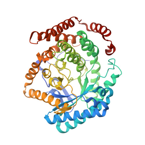
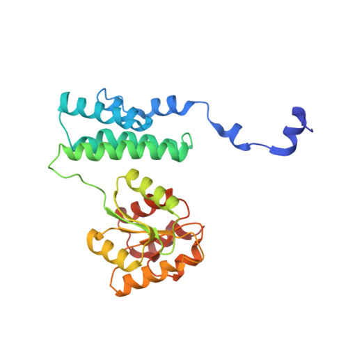
Insight into the mechanism of biological methanol activation based on the crystal structure of the methanol-cobalamin methyltransferase complex

Hagemeier, C.H.Krer, M.Thauer, R.K.Warkentin, E.Ermler, U.

(2006) Proc Natl Acad Sci U S A 103: 18917-18922

  • DOI: https://doi.org/10.1073/pnas.0603650103
  • Primary Citation of Related Structures:  
    2I2X

  • PubMed Abstract: 

    Some methanogenic and acetogenic microorganisms have the catalytic capability to cleave heterolytically the C O bond of methanol. To obtain insight into the elusive enzymatic mechanism of this challenging chemical reaction we have investigated the methanol-activating MtaBC complex from Methanosarcina barkeri composed of the zinc-containing MtaB and the 5-hydroxybenzimidazolylcobamide-carrying MtaC subunits. Here we report the 2.5-A crystal structure of this complex organized as a (MtaBC)(2) heterotetramer. MtaB folds as a TIM barrel and contains a novel zinc-binding motif. Zinc(II) lies at the bottom of a funnel formed at the C-terminal beta-barrel end and ligates to two cysteinyl sulfurs (Cys-220 and Cys-269) and one carboxylate oxygen (Glu-164). MtaC is structurally related to the cobalamin-binding domain of methionine synthase. Its corrinoid cofactor at the top of the Rossmann domain reaches deeply into the funnel of MtaB, defining a region between zinc(II) and the corrinoid cobalt that must be the binding site for methanol. The active site geometry supports a S(N)2 reaction mechanism, in which the C O bond in methanol is activated by the strong electrophile zinc(II) and cleaved because of an attack of the supernucleophile cob(I)amide. The environment of zinc(II) is characterized by an acidic cluster that increases the charge density on the zinc(II), polarizes methanol, and disfavors deprotonation of the methanol hydroxyl group. Implications of the MtaBC structure for the second step of the reaction, in which the methyl group is transferred to coenzyme M, are discussed.


  • Organizational Affiliation

    Max Planck Institute for Terrestrial Microbiology, Karl-von-Frisch-Strasse, D-35043 Marburg, Germany.


Macromolecules
Find similar proteins by:  (by identity cutoff)  |  3D Structure
Entity ID: 1
MoleculeChains Sequence LengthOrganismDetailsImage
Methyltransferase 1
A, C, E, G, I
A, C, E, G, I, K, M, O
461Methanosarcina barkeriMutation(s): 0 
EC: 2.1.1.90
UniProt
Find proteins for Q46EH3 (Methanosarcina barkeri (strain Fusaro / DSM 804))
Explore Q46EH3 
Go to UniProtKB:  Q46EH3
Entity Groups  
Sequence Clusters30% Identity50% Identity70% Identity90% Identity95% Identity100% Identity
UniProt GroupQ46EH3
Sequence Annotations
Expand
  • Reference Sequence
Find similar proteins by:  (by identity cutoff)  |  3D Structure
Entity ID: 2
MoleculeChains Sequence LengthOrganismDetailsImage
Methyltransferase 1
B, D, F, H, J
B, D, F, H, J, L, N, P
258Methanosarcina barkeriMutation(s): 0 
EC: 2.1.1.90
UniProt
Find proteins for Q46EH4 (Methanosarcina barkeri (strain Fusaro / DSM 804))
Explore Q46EH4 
Go to UniProtKB:  Q46EH4
Entity Groups  
Sequence Clusters30% Identity50% Identity70% Identity90% Identity95% Identity100% Identity
UniProt GroupQ46EH4
Sequence Annotations
Expand
  • Reference Sequence
Small Molecules
Ligands 3 Unique
IDChains Name / Formula / InChI Key2D Diagram3D Interactions
B13
Query on B13

Download Ideal Coordinates CCD File 
AA [auth F]
DA [auth H]
HA [auth J]
KA [auth L]
OA [auth N]
AA [auth F],
DA [auth H],
HA [auth J],
KA [auth L],
OA [auth N],
RA [auth P],
T [auth B],
W [auth D]
5-HYDROXYBENZIMIDAZOLYLCOB(III)AMIDE
C60 H88 Co N13 O15 P
QJVWXASLTDBQFK-LAYHTZHSSA-N
ZN
Query on ZN

Download Ideal Coordinates CCD File 
BA [auth G]
EA [auth I]
FA [auth I]
IA [auth K]
LA [auth M]
BA [auth G],
EA [auth I],
FA [auth I],
IA [auth K],
LA [auth M],
MA [auth M],
PA [auth O],
Q [auth A],
R [auth A],
U [auth C],
X [auth E],
Y [auth E]
ZINC ION
Zn
PTFCDOFLOPIGGS-UHFFFAOYSA-N
K
Query on K

Download Ideal Coordinates CCD File 
CA [auth G]
GA [auth I]
JA [auth K]
NA [auth M]
QA [auth O]
CA [auth G],
GA [auth I],
JA [auth K],
NA [auth M],
QA [auth O],
S [auth A],
V [auth C],
Z [auth E]
POTASSIUM ION
K
NPYPAHLBTDXSSS-UHFFFAOYSA-N
Experimental Data & Validation

Experimental Data

  • Method: X-RAY DIFFRACTION
  • Resolution: 2.50 Å
  • R-Value Free: 0.231 
  • R-Value Work: 0.182 
  • R-Value Observed: 0.184 
  • Space Group: P 1 21 1
Unit Cell:
Length ( Å )Angle ( ˚ )
a = 101.75α = 90
b = 172.85β = 98.86
c = 190.54γ = 90
Software Package:
Software NamePurpose
REFMACrefinement
ADSCdata collection
XDSdata reduction
XDSdata scaling
SHELXDphasing

Structure Validation

View Full Validation Report


Ligand Structure Quality Assessment 


Entry History 

Deposition Data

Revision History  (Full details and data files)

  • Version 1.0: 2006-11-21
    Type: Initial release
  • Version 1.1: 2008-04-21
    Changes: Version format compliance
  • Version 1.2: 2011-07-13
    Changes: Advisory, Derived calculations, Refinement description, Version format compliance
  • Version 1.3: 2024-02-21
    Changes: Data collection, Database references, Derived calculations, Refinement description