2I2F

Crystal structure of LmNADK1


Experimental Data Snapshot

  • Method: X-RAY DIFFRACTION
  • Resolution: 1.90 Å
  • R-Value Free: 0.243 
  • R-Value Work: 0.205 
  • R-Value Observed: 0.206 

wwPDB Validation   3D Report Full Report


Ligand Structure Quality Assessment 


This is version 1.4 of the entry. See complete history


Literature

NAD kinases use substrate-assisted catalysis for specific recognition of NAD.

Poncet-Montange, G.Assairi, L.Arold, S.Pochet, S.Labesse, G.

(2007) J Biol Chem 282: 33925-33934

  • DOI: https://doi.org/10.1074/jbc.M701394200
  • Primary Citation of Related Structures:  
    2I1W, 2I29, 2I2A, 2I2B, 2I2C, 2I2D, 2I2F, 2Q5F, 4DY6

  • PubMed Abstract: 

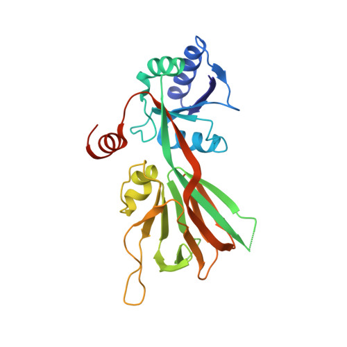
    Here we describe the crystal structures of the NAD kinase (LmNADK1) from Listeria monocytogenes in complex with its substrate NAD, its product NADP, or two synthesized NAD mimics. We identified one of the NAD mimics, di-adenosine diphosphate, as a new substrate for LmNADK1, whereas we showed that the closely related compound di-5'-thioadenosine is a novel non-natural inhibitor for this enzyme. These structures suggest a mechanism involving substrate-assisted catalysis. Indeed, sequence/structure comparison and directed mutagenesis have previously shown that NAD kinases (NADKs) and the distantly related 6-phosphofructokinases share the same catalytically important GGDGT motif. However, in this study we have shown that these enzymes use the central aspartate of this motif differently. Although this acidic residue chelates the catalytic Mg(2+) ion in 6-phosphofructokinases, it activates the phospho-acceptor (NAD) in NADKs. Sequence/structure comparisons suggest that the role of this aspartate would be conserved in NADKs and the related sphingosine and diacylglycerol kinases.


  • Organizational Affiliation

    Atelier de Bio- et Chimie Informatique Structurale, Centre de Biochimie Structurale, UMR 5048/CNRS, Universités Montpellier 1 et 2, 29 rue de Navacelles, Montpellier, France.


Macromolecules
Find similar proteins by:  (by identity cutoff)  |  3D Structure
Entity ID: 1
MoleculeChains Sequence LengthOrganismDetailsImage
Probable inorganic polyphosphate/ATP-NAD kinase 1272Listeria monocytogenesMutation(s): 1 
Gene Names: ppnK1
EC: 2.7.1.23
UniProt
Find proteins for Q8Y8D7 (Listeria monocytogenes serovar 1/2a (strain ATCC BAA-679 / EGD-e))
Explore Q8Y8D7 
Go to UniProtKB:  Q8Y8D7
Entity Groups  
Sequence Clusters30% Identity50% Identity70% Identity90% Identity95% Identity100% Identity
UniProt GroupQ8Y8D7
Sequence Annotations
Expand
  • Reference Sequence
Experimental Data & Validation

Experimental Data

  • Method: X-RAY DIFFRACTION
  • Resolution: 1.90 Å
  • R-Value Free: 0.243 
  • R-Value Work: 0.205 
  • R-Value Observed: 0.206 
  • Space Group: I 2 2 2
Unit Cell:
Length ( Å )Angle ( ˚ )
a = 62.428α = 90
b = 76.361β = 90
c = 119.202γ = 90
Software Package:
Software NamePurpose
REFMACrefinement
PDB_EXTRACTdata extraction
ADSCdata collection
MOSFLMdata reduction
SCALAdata scaling
MOLREPphasing

Structure Validation

View Full Validation Report


Ligand Structure Quality Assessment 


Entry History 

Deposition Data

Revision History  (Full details and data files)

  • Version 1.0: 2007-08-07
    Type: Initial release
  • Version 1.1: 2011-07-13
    Changes: Advisory, Derived calculations, Refinement description, Version format compliance
  • Version 1.2: 2018-02-14
    Changes: Experimental preparation
  • Version 1.3: 2021-10-20
    Changes: Database references, Derived calculations
  • Version 1.4: 2023-08-30
    Changes: Data collection, Refinement description