2HYI

Structure of the human exon junction complex with a trapped DEAD-box helicase bound to RNA


Experimental Data Snapshot

  • Method: X-RAY DIFFRACTION
  • Resolution: 2.30 Å
  • R-Value Free: 0.231 
  • R-Value Work: 0.212 
  • R-Value Observed: 0.213 

wwPDB Validation   3D Report Full Report


Ligand Structure Quality Assessment 


This is version 1.3 of the entry. See complete history


Literature

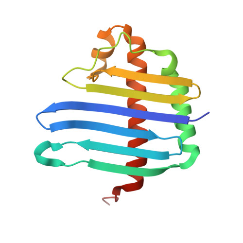
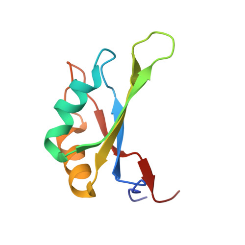
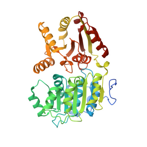
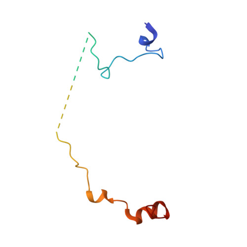
Structure of the exon junction core complex with a trapped DEAD-box ATPase bound to RNA.

Andersen, C.B.F.Ballut, L.Johansen, J.S.Chamieh, H.Nielsen, K.H.Oliveira, C.L.Pedersen, J.S.Seraphin, B.Le Hir, H.Andersen, G.R.

(2006) Science 313: 1968-1972

  • DOI: https://doi.org/10.1126/science.1131981
  • Primary Citation of Related Structures:  
    2HXY, 2HYI

  • PubMed Abstract: 

    In higher eukaryotes, a multiprotein exon junction complex is deposited on spliced messenger RNAs. The complex is organized around a stable core, which serves as a binding platform for numerous factors that influence messenger RNA function. Here, we present the crystal structure of a tetrameric exon junction core complex containing the DEAD-box adenosine triphosphatase (ATPase) eukaryotic initiation factor 4AIII (eIF4AIII) bound to an ATP analog, MAGOH, Y14, a fragment of MLN51, and a polyuracil mRNA mimic. eIF4AIII interacts with the phosphate-ribose backbone of six consecutive nucleotides and prevents part of the bound RNA from being double stranded. The MAGOH and Y14 subunits lock eIF4AIII in a prehydrolysis state, and activation of the ATPase probably requires only modest conformational changes in eIF4AIII motif I.


  • Organizational Affiliation

    Department of Molecular Biology, University of Aarhus, DK-8000 Aarhus, Denmark.


Macromolecules

Find similar proteins by:  (by identity cutoff)  |  3D Structure
Entity ID: 2
MoleculeChains Sequence LengthOrganismDetailsImage
Protein mago nashi homologC [auth A],
G
146Homo sapiensMutation(s): 0 
Gene Names: MAGOH
UniProt & NIH Common Fund Data Resources
Find proteins for P61326 (Homo sapiens)
Explore P61326 
Go to UniProtKB:  P61326
PHAROS:  P61326
GTEx:  ENSG00000162385 
Entity Groups  
Sequence Clusters30% Identity50% Identity70% Identity90% Identity95% Identity100% Identity
UniProt GroupP61326
Sequence Annotations
Expand
  • Reference Sequence
Find similar proteins by:  (by identity cutoff)  |  3D Structure
Entity ID: 3
MoleculeChains Sequence LengthOrganismDetailsImage
RNA-binding protein 8AD [auth B],
H
91Homo sapiensMutation(s): 0 
Gene Names: RBM8ARBM8
UniProt & NIH Common Fund Data Resources
Find proteins for Q9Y5S9 (Homo sapiens)
Explore Q9Y5S9 
Go to UniProtKB:  Q9Y5S9
PHAROS:  Q9Y5S9
GTEx:  ENSG00000265241 
Entity Groups  
Sequence Clusters30% Identity50% Identity70% Identity90% Identity95% Identity100% Identity
UniProt GroupQ9Y5S9
Sequence Annotations
Expand
  • Reference Sequence
Find similar proteins by:  (by identity cutoff)  |  3D Structure
Entity ID: 4
MoleculeChains Sequence LengthOrganismDetailsImage
Probable ATP-dependent RNA helicase DDX48E [auth C],
I
413Homo sapiensMutation(s): 0 
Gene Names: DDX48EIF4A3KIAA0111
EC: 3.6.1
UniProt & NIH Common Fund Data Resources
Find proteins for P38919 (Homo sapiens)
Explore P38919 
Go to UniProtKB:  P38919
PHAROS:  P38919
GTEx:  ENSG00000141543 
Entity Groups  
Sequence Clusters30% Identity50% Identity70% Identity90% Identity95% Identity100% Identity
UniProt GroupP38919
Sequence Annotations
Expand
  • Reference Sequence
Find similar proteins by:  (by identity cutoff)  |  3D Structure
Entity ID: 5
MoleculeChains Sequence LengthOrganismDetailsImage
Protein CASC3F [auth D],
J
77Homo sapiensMutation(s): 0 
Gene Names: CASC3MLN51
UniProt & NIH Common Fund Data Resources
Find proteins for O15234 (Homo sapiens)
Explore O15234 
Go to UniProtKB:  O15234
PHAROS:  O15234
GTEx:  ENSG00000108349 
Entity Groups  
Sequence Clusters30% Identity50% Identity70% Identity90% Identity95% Identity100% Identity
UniProt GroupO15234
Sequence Annotations
Expand
  • Reference Sequence

Find similar nucleic acids by:  Sequence   |   3D Structure  

Entity ID: 1
MoleculeChains LengthOrganismImage
5'-R(*UP*UP*UP*UP*UP*U)-3'A [auth F],
B [auth L]
6N/A
Sequence Annotations
Expand
  • Reference Sequence
Experimental Data & Validation

Experimental Data

  • Method: X-RAY DIFFRACTION
  • Resolution: 2.30 Å
  • R-Value Free: 0.231 
  • R-Value Work: 0.212 
  • R-Value Observed: 0.213 
  • Space Group: C 1 2 1
Unit Cell:
Length ( Å )Angle ( ˚ )
a = 185.83α = 90
b = 88.26β = 110.77
c = 145.77γ = 90
Software Package:
Software NamePurpose
PHASERphasing
CNSrefinement
XDSdata reduction
XSCALEdata scaling

Structure Validation

View Full Validation Report


Ligand Structure Quality Assessment 


Entry History 

Deposition Data

Revision History  (Full details and data files)

  • Version 1.0: 2006-08-15
    Type: Initial release
  • Version 1.1: 2008-05-01
    Changes: Version format compliance
  • Version 1.2: 2011-07-13
    Changes: Version format compliance
  • Version 1.3: 2024-02-21
    Changes: Data collection, Database references, Derived calculations