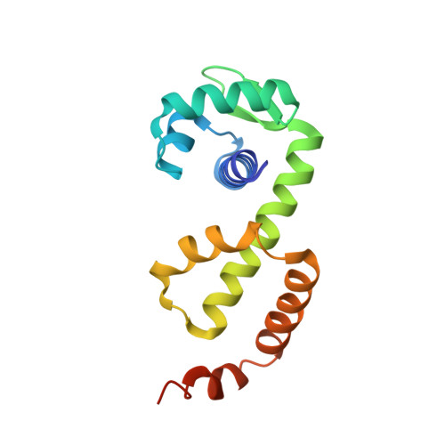
The conformations of the manganese transport regulator of Bacillus subtilis in its metal-free state.
DeWitt, M.A., Kliegman, J.I., Helmann, J.D., Brennan, R.G., Farrens, D.L., Glasfeld, A.(2007) J Mol Biol 365: 1257-1265
- PubMed: 17118401
- DOI: https://doi.org/10.1016/j.jmb.2006.10.080
- Primary Citation of Related Structures:
2HYF, 2HYG - PubMed Abstract:
The manganese transport regulator (MntR) from Bacillus subtilis binds cognate DNA sequences in response to elevated manganese concentrations. MntR functions as a homodimer that binds two manganese ions per subunit. Metal binding takes place at the interface of the two domains that comprise each MntR subunit: an N-terminal DNA-binding domain and a C-terminal dimerization domain. In order to elucidate the link between metal binding and activation, a crystallographic study of MntR in its metal-free state has been undertaken. Here we describe the structures of the native protein and a selenomethionine-containing variant, solved to 2.8 A. The two structures contain five crystallographically unique subunits of MntR, providing diverse views of the metal-free protein. In apo-MntR, as in the manganese complex, the dimer is formed by dyad-related C-terminal domains that provide a conserved structural core. Similarly, each DNA-binding domain largely retains the folded conformation found in metal bound forms of MntR. However, compared to metal-activated MntR, the DNA-binding domains move substantially with respect to the dimer interface in apo-MntR. Overlays of multiple apo-MntR structures indicate that there is a greater range of positioning allowed between N and C-terminal domains in the metal-free state and that the DNA-binding domains of the dimer are farther apart than in the activated complex. To further investigate the conformation of the DNA-binding domain of apo-MntR, a site-directed spin labeling experiment was performed on a mutant of MntR containing cysteine at residue 6. Consistent with the crystallographic results, EPR spectra of the spin-labeled mutant indicate that tertiary structure is conserved in the presence or absence of bound metals, though slightly greater flexibility is present in inactive forms of MntR.
Organizational Affiliation:
Department of Chemistry, Reed College, Portland, OR 97202, USA.