2HXZ

Crystal Structure of Cathepsin S in complex with a Nonpeptidic Inhibitor (Hexagonal spacegroup)


Experimental Data Snapshot

  • Method: X-RAY DIFFRACTION
  • Resolution: 1.90 Å
  • R-Value Free: 0.238 
  • R-Value Work: 0.219 
  • R-Value Observed: 0.220 

wwPDB Validation   3D Report Full Report


This is version 1.3 of the entry. See complete history


Literature

Identification of selective, nonpeptidic nitrile inhibitors of cathepsin s using the substrate activity screening method.

Patterson, A.W.Wood, W.J.Hornsby, M.Lesley, S.Spraggon, G.Ellman, J.A.

(2006) J Med Chem 49: 6298-6307

  • DOI: https://doi.org/10.1021/jm060701s
  • Primary Citation of Related Structures:  
    2H7J, 2HXZ

  • PubMed Abstract: 

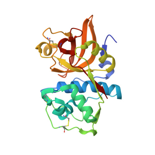
    The substrate activity screening method, a substrate-based fragment identification and optimization method for the development of enzyme inhibitors, was previously applied to cathepsin S to obtain low nanomolar 1,4-disubstituted-1,2,3-triazole-based aldehyde inhibitors (Wood, W. J. L.; Patterson, A. W.; Tsuruoka, H.; Jain, R. K.; Ellman, J. A. J. Am. Chem. Soc. 2005, 127, 15521-15527). Replacement of the metabolically labile aldehyde pharmacophore with the nitrile pharmacophore provided inhibitors with moderate potency for cathepsin S. The inhibitors showed good selectivity over cathepsins B and L but no selectivity over cathepsin K. X-ray structures of two crystal forms (1.5 and 1.9 A) of a complex between cathepsin S and a triazole inhibitor incorporating a chloromethyl ketone pharmacophore guided the design of triazole substrates with increased cleavage efficiency and selectivity for cathepsin S over cathepsins B, L, and K. Conversion of select substrates to nitrile inhibitors yielded a low molecular weight (414 Da) and potent (15 nM) cathepsin S inhibitor that showed >1000-fold selectivity over cathepsins B, L, and K.


  • Organizational Affiliation

    Department of Chemistry, University of California, Berkeley, California 94720, USA.


Macromolecules
Find similar proteins by:  (by identity cutoff)  |  3D Structure
Entity ID: 1
MoleculeChains Sequence LengthOrganismDetailsImage
Cathepsin S
A, B, C
220Homo sapiensMutation(s): 0 
Gene Names: CTSS
EC: 3.4.22.27
UniProt & NIH Common Fund Data Resources
Find proteins for P25774 (Homo sapiens)
Explore P25774 
Go to UniProtKB:  P25774
PHAROS:  P25774
GTEx:  ENSG00000163131 
Entity Groups  
Sequence Clusters30% Identity50% Identity70% Identity90% Identity95% Identity100% Identity
UniProt GroupP25774
Sequence Annotations
Expand
  • Reference Sequence
Small Molecules
Ligands 2 Unique
IDChains Name / Formula / InChI Key2D Diagram3D Interactions
H7J
Query on H7J

Download Ideal Coordinates CCD File 
E [auth A],
F [auth B],
G [auth C]
N-[(1S)-1-{1-[(1R,3E)-1-ACETYLPENT-3-EN-1-YL]-1H-1,2,3-TRIAZOL-4-YL}-1,2-DIMETHYLPROPYL]BENZAMIDE
C21 H28 N4 O2
OEHUTYXPQSSKAK-RVLLMHTFSA-N
SO4
Query on SO4

Download Ideal Coordinates CCD File 
D [auth A]SULFATE ION
O4 S
QAOWNCQODCNURD-UHFFFAOYSA-L
Experimental Data & Validation

Experimental Data

  • Method: X-RAY DIFFRACTION
  • Resolution: 1.90 Å
  • R-Value Free: 0.238 
  • R-Value Work: 0.219 
  • R-Value Observed: 0.220 
  • Space Group: P 65 2 2
Unit Cell:
Length ( Å )Angle ( ˚ )
a = 165.928α = 90
b = 165.928β = 90
c = 142.082γ = 120
Software Package:
Software NamePurpose
REFMACrefinement
HKL-2000data collection
HKL-2000data reduction
HKL-2000data scaling
MOLREPphasing

Structure Validation

View Full Validation Report


Entry History 

Deposition Data

Revision History  (Full details and data files)

  • Version 1.0: 2006-10-24
    Type: Initial release
  • Version 1.1: 2008-05-01
    Changes: Version format compliance
  • Version 1.2: 2011-07-13
    Changes: Version format compliance
  • Version 1.3: 2023-08-30
    Changes: Data collection, Database references, Derived calculations, Refinement description