2HWL

Crystal structure of thrombin in complex with fibrinogen gamma' peptide


Experimental Data Snapshot

  • Method: X-RAY DIFFRACTION
  • Resolution: 2.40 Å
  • R-Value Free: 0.249 
  • R-Value Work: 0.214 
  • R-Value Observed: 0.214 

wwPDB Validation   3D Report Full Report


Ligand Structure Quality Assessment 


This is version 1.6 of the entry. See complete history


Literature

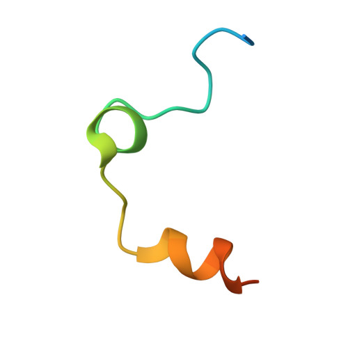
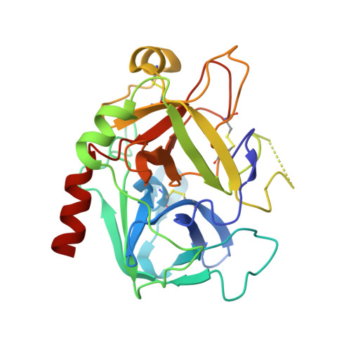
Crystal structure of thrombin in complex with fibrinogen gamma' peptide.

Pineda, A.O.Chen, Z.W.Marino, F.Mathews, F.S.Mosesson, M.W.Di Cera, E.

(2007) Biophys Chem 125: 556-559

  • DOI: https://doi.org/10.1016/j.bpc.2006.08.005
  • Primary Citation of Related Structures:  
    2HWL

  • PubMed Abstract: 

    Elevated levels of heterodimeric gamma(A)/gamma' fibrinogen 2 have been associated with an increased incidence of coronary artery disease, whereas a lowered content of gamma' chains is associated with an increased risk of venous thrombosis. Both situations may be related to the unique features of thrombin binding to variant gamma' chains. The gamma' peptide is an anionic fragment that binds thrombin with high affinity without interfering directly with substrate binding. Here we report the crystal structure of thrombin bound to the gamma' peptide, solved at 2.4 A resolution. The complex reveals extensive interactions between thrombin and the gamma' peptide mediated by electrostatic contacts with residues of exosite II and hydrophobic interactions with a pocket in close proximity to the Na(+) binding site. In its binding mode, the gamma' peptide completely overlaps with heparin bound to exosite II. These findings are consistent with functional data and broaden our understanding of how thrombin interacts with fibrinogen at the molecular level.


Macromolecules
Find similar proteins by:  (by identity cutoff)  |  3D Structure
Entity ID: 1
MoleculeChains Sequence LengthOrganismDetailsImage
Prothrombin
A, C
36Homo sapiensMutation(s): 0 
Gene Names: F2
EC: 3.4.21.5
UniProt & NIH Common Fund Data Resources
Find proteins for P00734 (Homo sapiens)
Explore P00734 
Go to UniProtKB:  P00734
PHAROS:  P00734
GTEx:  ENSG00000180210 
Entity Groups  
Sequence Clusters30% Identity50% Identity70% Identity90% Identity95% Identity100% Identity
UniProt GroupP00734
Sequence Annotations
Expand
  • Reference Sequence
Find similar proteins by:  (by identity cutoff)  |  3D Structure
Entity ID: 2
MoleculeChains Sequence LengthOrganismDetailsImage
Prothrombin
B, D
259Homo sapiensMutation(s): 1 
Gene Names: F2
EC: 3.4.21.5
UniProt & NIH Common Fund Data Resources
Find proteins for P00734 (Homo sapiens)
Explore P00734 
Go to UniProtKB:  P00734
PHAROS:  P00734
GTEx:  ENSG00000180210 
Entity Groups  
Sequence Clusters30% Identity50% Identity70% Identity90% Identity95% Identity100% Identity
UniProt GroupP00734
Sequence Annotations
Expand
  • Reference Sequence

Find similar proteins by:  Sequence   |   3D Structure  

Entity ID: 3
MoleculeChains Sequence LengthOrganismDetailsImage
Fibrinogen gamma' peptideE [auth P]14N/AMutation(s): 2 
UniProt & NIH Common Fund Data Resources
Find proteins for P02679 (Homo sapiens)
Explore P02679 
Go to UniProtKB:  P02679
PHAROS:  P02679
GTEx:  ENSG00000171557 
Entity Groups  
Sequence Clusters30% Identity50% Identity70% Identity90% Identity95% Identity100% Identity
UniProt GroupP02679
Sequence Annotations
Expand
  • Reference Sequence
Small Molecules
Modified Residues  1 Unique
IDChains TypeFormula2D DiagramParent
PTR
Query on PTR
E [auth P]L-PEPTIDE LINKINGC9 H12 N O6 PTYR
Experimental Data & Validation

Experimental Data

  • Method: X-RAY DIFFRACTION
  • Resolution: 2.40 Å
  • R-Value Free: 0.249 
  • R-Value Work: 0.214 
  • R-Value Observed: 0.214 
  • Space Group: P 1 21 1
Unit Cell:
Length ( Å )Angle ( ˚ )
a = 66.397α = 90
b = 81.523β = 109.35
c = 70.256γ = 90
Software Package:
Software NamePurpose
CNSrefinement
HKL-2000data collection
HKL-2000data reduction
HKL-2000data scaling
CNSphasing

Structure Validation

View Full Validation Report


Ligand Structure Quality Assessment 


Entry History 

Deposition Data

Revision History  (Full details and data files)

  • Version 1.0: 2006-09-19
    Type: Initial release
  • Version 1.1: 2008-05-01
    Changes: Version format compliance
  • Version 1.2: 2011-07-13
    Changes: Non-polymer description, Version format compliance
  • Version 1.3: 2020-07-29
    Type: Remediation
    Reason: Carbohydrate remediation
    Changes: Data collection, Database references, Derived calculations, Structure summary
  • Version 1.4: 2021-10-20
    Changes: Database references, Structure summary
  • Version 1.5: 2023-08-30
    Changes: Data collection, Refinement description
  • Version 1.6: 2023-11-15
    Changes: Data collection