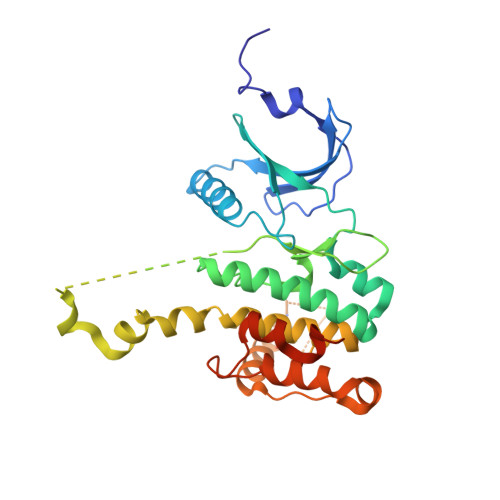
2HW7

Crystal Structure of Mnk2-D228G in complex with Staurosporine


Experimental Data Snapshot

  • Method: X-RAY DIFFRACTION
  • Resolution: 2.71 Å
  • R-Value Free: 0.246 
  • R-Value Work: 0.201 
  • R-Value Observed: 0.203 

wwPDB Validation   3D Report Full Report


Ligand Structure Quality Assessment 


This is version 1.4 of the entry. See complete history


Literature

Mitogen-activated protein kinases interacting kinases are autoinhibited by a reprogrammed activation segment.

Jauch, R.Cho, M.K.Netter, C.Schreiter, K.Aicher, B.Zweckstetter, M.Wahl, M.C.

(2006) EMBO J 25: 4020-4032

  • DOI: https://doi.org/10.1038/sj.emboj.7601285
  • Primary Citation of Related Structures:  
    2HW6, 2HW7

  • PubMed Abstract: 

    Autoinhibition is a recurring mode of protein kinase regulation and can be based on diverse molecular mechanisms. Here, we show by crystal structure analysis, nuclear magnetic resonance (NMR)-based nucleotide affinity studies and rational mutagenesis that nonphosphorylated mitogen-activated protein (MAP) kinases interacting kinase (Mnk) 1 is autoinhibited by conversion of the activation segment into an autoinhibitory module. In a Mnk1 crystal structure, the activation segment is repositioned via a Mnk-specific sequence insertion at the N-terminal lobe with the following consequences: (i) the peptide substrate binding site is deconstructed, (ii) the interlobal cleft is narrowed, (iii) an essential Lys-Glu pair is disrupted and (iv) the magnesium-binding loop is locked into an ATP-competitive conformation. Consistently, deletion of the Mnk-specific insertion or removal of a conserved phenylalanine side chain, which induces a blockade of the ATP pocket, increase the ATP affinity of Mnk1. Structural rearrangements required for the activation of Mnks are apparent from the cocrystal structure of a Mnk2 D228G -staurosporine complex and can be modeled on the basis of crystal packing interactions. Our data suggest a novel regulatory mechanism specific for the Mnk subfamily.


  • Organizational Affiliation

    Max-Planck-Institut für Biophysikalische Chemie, Abteilung Molekulare Entwicklungsbiologie, Göttingen, Germany. jauchr@gis.a-star.edu.sg


Macromolecules
Find similar proteins by:  (by identity cutoff)  |  3D Structure
Entity ID: 1
MoleculeChains Sequence LengthOrganismDetailsImage
MAP kinase-interacting serine/threonine-protein kinase 2316Homo sapiensMutation(s): 1 
Gene Names: MKNK2MNK2
EC: 2.7.11.1
UniProt & NIH Common Fund Data Resources
Find proteins for Q9HBH9 (Homo sapiens)
Explore Q9HBH9 
Go to UniProtKB:  Q9HBH9
PHAROS:  Q9HBH9
GTEx:  ENSG00000099875 
Entity Groups  
Sequence Clusters30% Identity50% Identity70% Identity90% Identity95% Identity100% Identity
UniProt GroupQ9HBH9
Sequence Annotations
Expand
  • Reference Sequence
Small Molecules
Experimental Data & Validation

Experimental Data

  • Method: X-RAY DIFFRACTION
  • Resolution: 2.71 Å
  • R-Value Free: 0.246 
  • R-Value Work: 0.201 
  • R-Value Observed: 0.203 
  • Space Group: P 32 2 1
Unit Cell:
Length ( Å )Angle ( ˚ )
a = 102.366α = 90
b = 102.366β = 90
c = 76.439γ = 120
Software Package:
Software NamePurpose
REFMACrefinement
HKL-2000data reduction
HKL-2000data scaling
MOLREPphasing

Structure Validation

View Full Validation Report


Ligand Structure Quality Assessment 


Entry History 

Deposition Data

Revision History  (Full details and data files)

  • Version 1.0: 2006-08-29
    Type: Initial release
  • Version 1.1: 2008-05-01
    Changes: Version format compliance
  • Version 1.2: 2011-07-13
    Changes: Advisory, Version format compliance
  • Version 1.3: 2021-10-20
    Changes: Database references, Derived calculations
  • Version 1.4: 2023-08-30
    Changes: Data collection, Refinement description