The Acylaminoacyl Peptidase from Aeropyrum pernix K1 Thought to Be an Exopeptidase Displays Endopeptidase Activity
Kiss, A.L., Hornung, B., Radi, K., Gengeliczki, Z., Sztaray, B., Juhasz, T., Szeltner, Z., Harmat, V., Polgar, L.(2007) J Mol Biol 368: 509-520
- PubMed: 17350041
- DOI: https://doi.org/10.1016/j.jmb.2007.02.025
- Primary Citation of Related Structures:
2HU5, 2HU7, 2HU8 - PubMed Abstract:
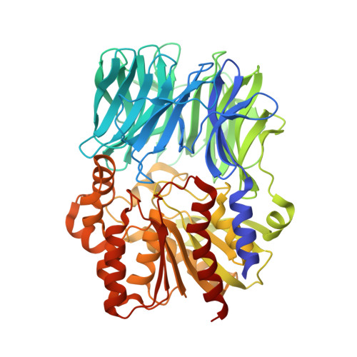
Mammalian acylaminoacyl peptidase, a member of the prolyl oligopeptidase family of serine peptidases, is an exopeptidase, which removes acylated amino acid residues from the N terminus of oligopeptides. We have investigated the kinetics and inhibitor binding of the orthologous acylaminoacyl peptidase from the thermophile Aeropyrum pernix K1 (ApAAP). Complex pH-rate profiles were found with charged substrates, indicating a strong electrostatic effect in the surroundings of the active site. Unexpectedly, we have found that oligopeptides can be hydrolysed beyond the N-terminal peptide bond, demonstrating that ApAAP exhibits endopeptidase activity. It was thought that the enzyme is specific for hydrophobic amino acids, in particular phenylalanine, in accord with the non-polar S1 subsite of ApAAP. However, cleavage after an Ala residue contradicted this notion and demonstrated that P1 residues of different nature may bind to the S1 subsite depending on the remaining peptide residues. The crystal structures of the complexes formed between the enzyme and product-like inhibitors identified the oxyanion-binding site unambiguously and demonstrated that the phenylalanine ring of the P1 peptide residue assumes a position different from that established in a previous study, using 4-nitrophenylphosphate. We have found that the substrate-binding site extends beyond the S2 subsite, being capable of binding peptides with a longer N terminus. The S2 subsite displays a non-polar character, which is unique among the enzymes of this family. The S3 site was identified as a hydrophobic region that does not form hydrogen bonds with the inhibitor P3 residue. The enzyme-inhibitor complexes revealed that, upon ligand-binding, the S1 subsite undergoes significant conformational changes, demonstrating the plasticity of the specificity site.
Organizational Affiliation:
Institute of Enzymology, Biological Research Center, Hungarian Academy of Sciences, H-1518 Budapest 112, P.O. Box 7, Hungary.