Methylene substitution at the alpha-beta bridging position within the phosphate chain of dUDP profoundly perturbs ligand accommodation into the dUTPase active site.
Kovari, J., Barabas, O., Varga, B., Bekesi, A., Tolgyesi, F., Fidy, J., Nagy, J., Vertessy, B.G.(2008) Proteins 71: 308-319
- PubMed: 17932923
- DOI: https://doi.org/10.1002/prot.21757
- Primary Citation of Related Structures:
2HR6, 2HRM - PubMed Abstract:
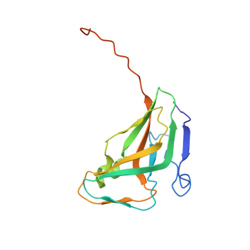
dUTP pyrophosphatase, a preventive DNA repair enzyme, contributes to maintain the appropriate cellular dUTP/dTTP ratio by catalyzing dUTP hydrolysis. dUTPase is essential for viability in bacteria and eukaryotes alike. Identification of species-specific antagonists of bacterial dUTPases is expected to contribute to the development of novel antimicrobial agents. As a first general step, design of dUTPase inhibitors should be based on modifications of the substrate dUTP phosphate chain, as modifications in either base or sugar moieties strongly impair ligand binding. Based on structural differences between bacterial and human dUTPases, derivatization of dUTP-analogous compounds will be required as a second step to invoke species-specific character. Studies performed with dUTP analogues also offer insights into substrate binding characteristics of this important and structurally peculiar enzyme. In this study, alpha,beta-methylene-dUDP was synthesized and its complex with dUTPase was characterized. Enzymatic phosphorylation of this substrate analogue by pyruvate kinase was not possible in contrast to the successful enzymatic phosphorylation of alpha,beta-imino-dUDP. One explanation for this finding is that the different bond angles and the presence of the methylene group may preclude formation of a catalytically competent complex with the kinase. Crystal structure of E. coli dUTPase:alpha,beta-methylene-dUDP and E. coli dUTPase:dUDP:Mn complexes were determined and analyzed in comparison with previous data. Results show that the "trans" alpha-phosphate conformation of alpha,beta-methylene-dUDP differs from the catalytically competent "gauche" alpha-phosphate conformation of the imino analogue and the oxo substrate, manifested in the shifted position of the alpha-phosphorus by more than 3 A. The three-dimensional structures determined in this work show that the binding of the methylene analogue with the alpha-phosphorus in the "gauche" conformation would result in steric clash of the methylene group with the protein atoms. In addition, the metal ion cofactor was not bound in the crystal of the complex with the methylene analogue while it was clearly visible as coordinated to dUDP, arguing that the altered phosphate chain conformation also perturbs metal ion complexation. Isothermal calorimetry titrations indicate that the binding affinity of alpha,beta-methylene-dUDP toward dUTPase is drastically decreased when compared with that of dUDP. In conclusion, the present data suggest that while alpha,beta-methylene-dUDP seems to be practically nonhydrolyzable, it is not a strong binding inhibitor of dUTPase probably due to the altered binding mode of the phosphate chain. Results indicate that in some cases methylene analogues may not faithfully reflect the competent substrate ligand properties, especially if the methylene hydrogens are in steric conflict with the protein.
Organizational Affiliation:
Institute of Enzymology, Biological Research Center, Hungarian Academy of Sciences, Budapest, Hungary.