2HQS

Crystal structure of TolB/Pal complex


Experimental Data Snapshot

  • Method: X-RAY DIFFRACTION
  • Resolution: 1.50 Å
  • R-Value Free: 0.213 
  • R-Value Work: 0.180 
  • R-Value Observed: 0.182 

wwPDB Validation   3D Report Full Report


This is version 1.3 of the entry. See complete history


Literature

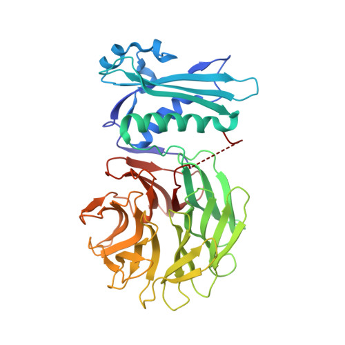
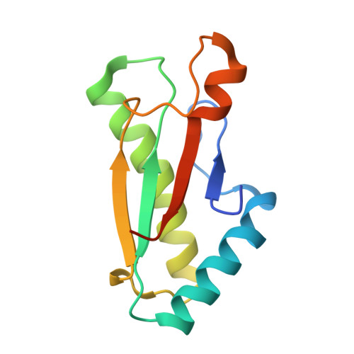
Molecular mimicry enables competitive recruitment by a natively disordered protein.

Bonsor, D.A.Grishkovskaya, I.Dodson, E.J.Kleanthous, C.

(2007) J Am Chem Soc 129: 4800-4807

  • DOI: https://doi.org/10.1021/ja070153n
  • Primary Citation of Related Structures:  
    2HQS

  • PubMed Abstract: 

    We report the crystal structure of the Escherichia coli TolB-Pal complex, a protein-protein complex involved in maintaining the integrity of the outer membrane (OM) in all Gram-negative bacteria that is parasitized by colicins (protein antibiotics) to expedite their entry into cells. Nuclease colicins competitively recruit TolB using their natively disordered regions (NDRs) to disrupt its complex with Pal, which is thought to trigger translocation of the toxin across a locally destabilized OM. The structure shows induced-fit binding of peptidoglycan-associated lipoprotein (Pal) to the beta-propeller domain of TolB causing the N-terminus of one of its alpha-helices to unwind and several residues to undergo substantial changes in conformation. The resulting interactions with TolB are known to be essential for the stability of the complex and the bacterial OM. Structural comparisons with a TolB-colicin NDR complex reveal that colicins bind at the Pal site, mimicking rearranged Pal residues while simultaneously appearing to block induced-fit changes in TolB. The study therefore explains how colicins recruit TolB in the bacterial periplasm and highlights a novel binding mechanism for a natively disordered protein.


  • Organizational Affiliation

    Department of Biology, University of York, Heslington, York, YO10 5YW, United Kingdom.


Macromolecules
Find similar proteins by:  (by identity cutoff)  |  3D Structure
Entity ID: 1
MoleculeChains Sequence LengthOrganismDetailsImage
Protein tolBA,
C [auth B],
E [auth D],
G [auth F]
415Escherichia coliMutation(s): 0 
Gene Names: tolB
UniProt
Find proteins for P0A855 (Escherichia coli (strain K12))
Explore P0A855 
Go to UniProtKB:  P0A855
Entity Groups  
Sequence Clusters30% Identity50% Identity70% Identity90% Identity95% Identity100% Identity
UniProt GroupP0A855
Sequence Annotations
Expand
  • Reference Sequence
Find similar proteins by:  (by identity cutoff)  |  3D Structure
Entity ID: 2
MoleculeChains Sequence LengthOrganismDetailsImage
Peptidoglycan-associated lipoproteinB [auth H],
D [auth C],
F [auth E],
H [auth G]
118Escherichia coliMutation(s): 0 
Gene Names: palexcC
UniProt
Find proteins for P0A912 (Escherichia coli (strain K12))
Explore P0A912 
Go to UniProtKB:  P0A912
Entity Groups  
Sequence Clusters30% Identity50% Identity70% Identity90% Identity95% Identity100% Identity
UniProt GroupP0A912
Sequence Annotations
Expand
  • Reference Sequence
Small Molecules
Ligands 3 Unique
IDChains Name / Formula / InChI Key2D Diagram3D Interactions
SO4
Query on SO4

Download Ideal Coordinates CCD File 
J [auth A]
O [auth B]
S [auth D]
W [auth F]
X [auth F]
J [auth A],
O [auth B],
S [auth D],
W [auth F],
X [auth F],
Y [auth F]
SULFATE ION
O4 S
QAOWNCQODCNURD-UHFFFAOYSA-L
GOL
Query on GOL

Download Ideal Coordinates CCD File 
AA [auth F]
K [auth A]
L [auth A]
P [auth B]
Q [auth B]
AA [auth F],
K [auth A],
L [auth A],
P [auth B],
Q [auth B],
T [auth D],
Z [auth F]
GLYCEROL
C3 H8 O3
PEDCQBHIVMGVHV-UHFFFAOYSA-N
ACT
Query on ACT

Download Ideal Coordinates CCD File 
BA [auth G]
I [auth A]
M [auth H]
N [auth B]
R [auth C]
BA [auth G],
I [auth A],
M [auth H],
N [auth B],
R [auth C],
U [auth E],
V [auth F]
ACETATE ION
C2 H3 O2
QTBSBXVTEAMEQO-UHFFFAOYSA-M
Experimental Data & Validation

Experimental Data

  • Method: X-RAY DIFFRACTION
  • Resolution: 1.50 Å
  • R-Value Free: 0.213 
  • R-Value Work: 0.180 
  • R-Value Observed: 0.182 
  • Space Group: P 1
Unit Cell:
Length ( Å )Angle ( ˚ )
a = 74.847α = 87.15
b = 89.049β = 89.62
c = 91.051γ = 68.81
Software Package:
Software NamePurpose
REFMACrefinement
MOSFLMdata reduction
CCP4data scaling
MOLREPphasing

Structure Validation

View Full Validation Report


Entry History 

Deposition Data

Revision History  (Full details and data files)

  • Version 1.0: 2007-04-03
    Type: Initial release
  • Version 1.1: 2008-05-01
    Changes: Version format compliance
  • Version 1.2: 2011-07-13
    Changes: Non-polymer description, Version format compliance
  • Version 1.3: 2022-12-21
    Changes: Database references, Derived calculations