2HPO

Structure of Aminopeptidase N from E. coli Suggests a Compartmentalized, Gated Active Site


Experimental Data Snapshot

  • Method: X-RAY DIFFRACTION
  • Resolution: 1.65 Å
  • R-Value Free: 0.181 
  • R-Value Work: 0.157 
  • R-Value Observed: 0.158 

wwPDB Validation   3D Report Full Report


This is version 1.3 of the entry. See complete history


Literature

Structure of aminopeptidase N from Escherichia coli suggests a compartmentalized, gated active site.

Addlagatta, A.Gay, L.Matthews, B.W.

(2006) Proc Natl Acad Sci U S A 103: 13339-13344

  • DOI: https://doi.org/10.1073/pnas.0606167103
  • Primary Citation of Related Structures:  
    2HPO, 2HPT

  • PubMed Abstract: 

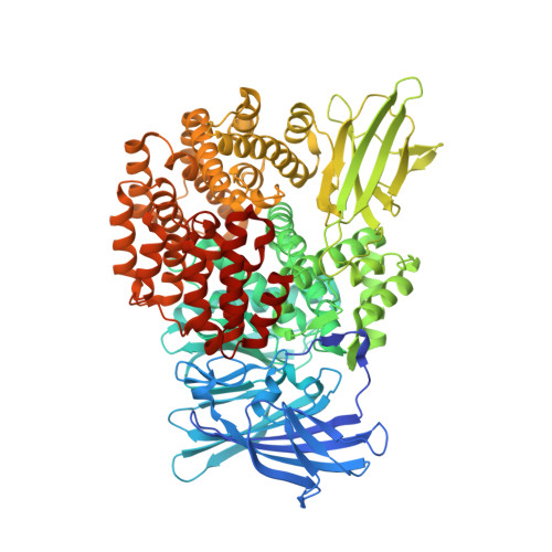
    Aminopeptidase N from Escherichia coli is a major metalloprotease that participates in the controlled hydrolysis of peptides in the proteolytic pathway. Determination of the 870-aa structure reveals that it has four domains similar to the tricorn-interacting factor F3. The thermolysin-like active site is enclosed within a large cavity with a volume of 2,200 A(3), which is inaccessible to substrates except for a small opening of approximately 8-10 A. The substrate-based inhibitor bestatin binds to the protein with minimal changes, suggesting that this is the active form of the enzyme. The previously described structure of F3 had three distinct conformations that were described as "closed," "intermediate," and "open." The structure of aminopeptidase N from E. coli, however, is substantially more closed than any of these. Taken together, the results suggest that these proteases, which are involved in intracellular peptide degradation, prevent inadvertent hydrolysis of inappropriate substrates by enclosing the active site within a large cavity. There is also some evidence that the open form of the enzyme, which admits substrates, remains inactive until it adopts the closed form.


  • Organizational Affiliation

    Howard Hughes Medical Institute and Institute of Molecular Biology and Department of Physics, University of Oregon, Eugene, OR 97403-1229.


Macromolecules
Find similar proteins by:  (by identity cutoff)  |  3D Structure
Entity ID: 1
MoleculeChains Sequence LengthOrganismDetailsImage
Aminopeptidase N891Escherichia coli K-12Mutation(s): 0 
Gene Names: pepN
EC: 3.4.11.2
UniProt
Find proteins for P04825 (Escherichia coli (strain K12))
Explore P04825 
Go to UniProtKB:  P04825
Entity Groups  
Sequence Clusters30% Identity50% Identity70% Identity90% Identity95% Identity100% Identity
UniProt GroupP04825
Sequence Annotations
Expand
  • Reference Sequence
Experimental Data & Validation

Experimental Data

  • Method: X-RAY DIFFRACTION
  • Resolution: 1.65 Å
  • R-Value Free: 0.181 
  • R-Value Work: 0.157 
  • R-Value Observed: 0.158 
  • Space Group: P 31 2 1
Unit Cell:
Length ( Å )Angle ( ˚ )
a = 120.438α = 90
b = 120.438β = 90
c = 170.561γ = 120
Software Package:
Software NamePurpose
REFMACrefinement
HKL-2000data reduction
SCALEPACKdata scaling
SHELXSphasing

Structure Validation

View Full Validation Report


Entry History 

Deposition Data

Revision History  (Full details and data files)

  • Version 1.0: 2006-08-15
    Type: Initial release
  • Version 1.1: 2008-05-01
    Changes: Version format compliance
  • Version 1.2: 2011-07-13
    Changes: Non-polymer description, Version format compliance
  • Version 1.3: 2024-02-14
    Changes: Data collection, Database references, Derived calculations