2HMU

Diamond-shaped octameric ring structure of an RCK domain with ATP bound


Experimental Data Snapshot

  • Method: X-RAY DIFFRACTION
  • Resolution: 2.25 Å
  • R-Value Free: 0.245 
  • R-Value Work: 0.228 

wwPDB Validation   3D Report Full Report


Ligand Structure Quality Assessment 


This is version 1.5 of the entry. See complete history


Literature

The RCK Domain of the KtrAB K(+) Transporter: Multiple Conformations of an Octameric Ring.

Albright, R.A.Ibar, J.L.Kim, C.U.Gruner, S.M.Morais-Cabral, J.H.

(2006) Cell 126: 1147-1159

  • DOI: https://doi.org/10.1016/j.cell.2006.08.028
  • Primary Citation of Related Structures:  
    2HMS, 2HMT, 2HMU, 2HMV, 2HMW

  • PubMed Abstract: 

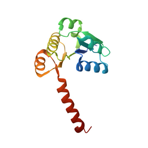
    The KtrAB ion transporter is a complex of the KtrB membrane protein and KtrA, an RCK domain. RCK domains regulate eukaryotic and prokaryotic membrane proteins involved in K(+) transport. Conflicting functional models have proposed two different oligomeric arrangements for RCK domains, tetramer versus octamer. Our results for the KtrAB RCK domain clearly show an octamer in solution and in the crystal. We determined the structure of this protein in three different octameric ring conformations that resemble the RCK-domain octamer observed in the MthK potassium channel but show striking differences in size and symmetry. We present experimental evidence for the association between one RCK octameric ring and two KtrB membrane proteins. These results provide insights into the quaternary organization of the KtrAB transporter and its mechanism of activation and show that the RCK-domain octameric ring model is generally applicable to other ion-transport systems.


  • Organizational Affiliation

    Department of Molecular Biophysics and Biochemistry, Yale University, 266 Whitney Avenue, New Haven, CT 06520, USA.


Macromolecules
Find similar proteins by:  (by identity cutoff)  |  3D Structure
Entity ID: 1
MoleculeChains Sequence LengthOrganismDetailsImage
YuaA protein
A, B
144Bacillus subtilisMutation(s): 1 
Gene Names: yuaA
UniProt
Find proteins for O32080 (Bacillus subtilis (strain 168))
Explore O32080 
Go to UniProtKB:  O32080
Entity Groups  
Sequence Clusters30% Identity50% Identity70% Identity90% Identity95% Identity100% Identity
UniProt GroupO32080
Sequence Annotations
Expand
  • Reference Sequence
Small Molecules
Ligands 1 Unique
IDChains Name / Formula / InChI Key2D Diagram3D Interactions
ATP
Query on ATP

Download Ideal Coordinates CCD File 
C [auth A],
D [auth B]
ADENOSINE-5'-TRIPHOSPHATE
C10 H16 N5 O13 P3
ZKHQWZAMYRWXGA-KQYNXXCUSA-N
Binding Affinity Annotations 
IDSourceBinding Affinity
ATP PDBBind:  2HMU Kd: 20 (nM) from 1 assay(s)
Experimental Data & Validation

Experimental Data

  • Method: X-RAY DIFFRACTION
  • Resolution: 2.25 Å
  • R-Value Free: 0.245 
  • R-Value Work: 0.228 
  • Space Group: P 4 2 2
Unit Cell:
Length ( Å )Angle ( ˚ )
a = 127.668α = 90
b = 127.668β = 90
c = 51.88γ = 90
Software Package:
Software NamePurpose
DENZOdata reduction
SCALEPACKdata scaling
CNSrefinement
PDB_EXTRACTdata extraction
HKL-2000data reduction
HKL-2000data scaling
MOLREPphasing

Structure Validation

View Full Validation Report


Ligand Structure Quality Assessment 


Entry History 

Deposition Data

Revision History  (Full details and data files)

  • Version 1.0: 2006-09-26
    Type: Initial release
  • Version 1.1: 2008-05-01
    Changes: Version format compliance
  • Version 1.2: 2011-07-13
    Changes: Version format compliance
  • Version 1.3: 2017-10-18
    Changes: Refinement description
  • Version 1.4: 2021-10-20
    Changes: Database references, Derived calculations
  • Version 1.5: 2023-08-30
    Changes: Data collection, Refinement description