2HH7

Crystal Structure of Cu(I) bound CsoR from Mycobacterium tuberculosis.


Experimental Data Snapshot

  • Method: X-RAY DIFFRACTION
  • Resolution: 2.55 Å
  • R-Value Free: 0.279 
  • R-Value Work: 0.231 

wwPDB Validation   3D Report Full Report


This is version 1.3 of the entry. See complete history


Literature

CsoR is a novel Mycobacterium tuberculosis copper-sensing transcriptional regulator.

Liu, T.Ramesh, A.Ma, Z.Ward, S.K.Zhang, L.George, G.N.Talaat, A.M.Sacchettini, J.C.Giedroc, D.P.

(2007) Nat Chem Biol 3: 60-68

  • DOI: https://doi.org/10.1038/nchembio844
  • Primary Citation of Related Structures:  
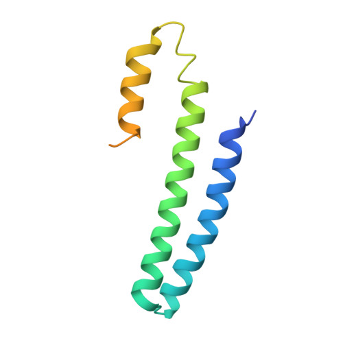
    2HH7

  • PubMed Abstract: 

    Copper is an essential element that becomes highly cytotoxic when concentrations exceed the capacity of cells to sequester the ion. Here, we identify a new copper-specific repressor (CsoR) of a copper-sensitive operon (cso) in Mycobacterium tuberculosis (Mtb) that is representative of a large, previously uncharacterized family of proteins (DUF156). Electronic and X-ray absorption spectroscopies reveal that CsoR binds a single-monomer mole equivalent of Cu(I) to form a trigonally coordinated (S(2)N) Cu(I) complex. The 2.6-A crystal structure of copper-loaded CsoR shows a homodimeric antiparallel four-helix bundle architecture that represents a novel DNA-binding fold. The Cu(I) is coordinated by Cys36, Cys65' and His61' in a subunit bridging site. Cu(I) binding negatively regulates the binding of CsoR to a DNA fragment encompassing the operator-promoter region of the Mtb cso operon; this results in derepression of the operon in Mtb and the heterologous host Mycobacterium smegmatis. Substitution of Cys36 or His61 with alanine abolishes Cu(I)- and CsoR-dependent regulation in vivo and in vitro. Potential roles of CsoR in Mtb pathogenesis are discussed.


  • Organizational Affiliation

    Department of Biochemistry and Biophysics, Texas A&M University, College Station, Texas 77843-2128, USA.


Macromolecules
Find similar proteins by:  (by identity cutoff)  |  3D Structure
Entity ID: 1
MoleculeChains Sequence LengthOrganismDetailsImage
Hypothetical protein CsoR119Mycobacterium tuberculosisMutation(s): 0 
UniProt
Find proteins for P9WP49 (Mycobacterium tuberculosis (strain ATCC 25618 / H37Rv))
Explore P9WP49 
Go to UniProtKB:  P9WP49
Entity Groups  
Sequence Clusters30% Identity50% Identity70% Identity90% Identity95% Identity100% Identity
UniProt GroupP9WP49
Sequence Annotations
Expand
  • Reference Sequence
Small Molecules
Ligands 1 Unique
IDChains Name / Formula / InChI Key2D Diagram3D Interactions
CU1
Query on CU1

Download Ideal Coordinates CCD File 
B [auth A]COPPER (I) ION
Cu
VMQMZMRVKUZKQL-UHFFFAOYSA-N
Experimental Data & Validation

Experimental Data

  • Method: X-RAY DIFFRACTION
  • Resolution: 2.55 Å
  • R-Value Free: 0.279 
  • R-Value Work: 0.231 
  • Space Group: P 64 2 2
Unit Cell:
Length ( Å )Angle ( ˚ )
a = 91.059α = 90
b = 91.059β = 90
c = 46.779γ = 120
Software Package:
Software NamePurpose
SOLVEphasing
PHENIXrefinement
HKL-2000data reduction
SCALEPACKdata scaling

Structure Validation

View Full Validation Report


Entry History 

Deposition Data

Revision History  (Full details and data files)

  • Version 1.0: 2006-12-05
    Type: Initial release
  • Version 1.1: 2008-05-01
    Changes: Version format compliance
  • Version 1.2: 2011-07-13
    Changes: Derived calculations, Version format compliance
  • Version 1.3: 2024-02-14
    Changes: Data collection, Database references, Derived calculations