Human Mitochondrial Branched Chain Aminotransferase Isozyme: STRUCTURAL ROLE OF THE CXXC CENTER IN CATALYSIS.
Yennawar, N.H., Islam, M.M., Conway, M., Wallin, R., Hutson, S.M.(2006) J Biol Chem 281: 39660-39671
- PubMed: 17050531
- DOI: https://doi.org/10.1074/jbc.M607552200
- Primary Citation of Related Structures:
2HDK, 2HG8, 2HGW, 2HGX, 2HHF - PubMed Abstract:
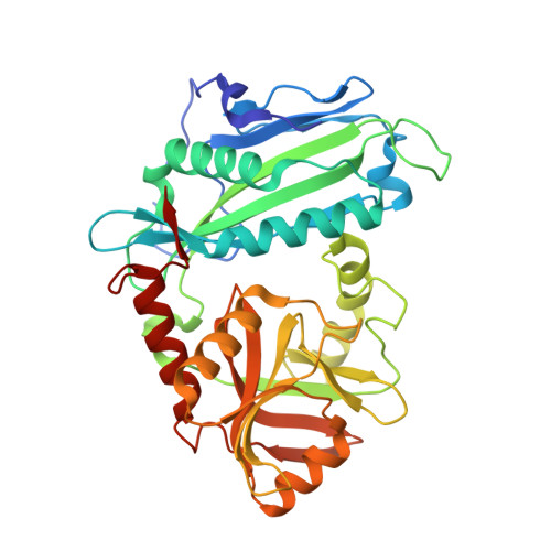
Mammalian branched chain aminotransferases (BCATs) have a unique CXXC center. Kinetic and structural studies of three CXXC center mutants (C315A, C318A, and C315A/C318A) of human mitochondrial (hBCATm) isozyme and the oxidized hBCATm enzyme (hBCATm-Ox) have been used to elucidate the role of this center in hBCATm catalysis. X-ray crystallography revealed that the CXXC motif, through its network of hydrogen bonds, plays a crucial role in orienting the substrate optimally for catalysis. In all structures, there were changes in the structure of the beta-turn preceding the CXXC motif when compared with wild type protein. The N-terminal loop between residues 15 and 32 is flexible in the oxidized and mutant enzymes, the disorder greater in the oxidized protein. Disordering of the N-terminal loop disrupts the integrity of the side chain binding pocket, particularly for the branched chain side chain, less so for the dicarboxylate substrate side chain. The kinetic studies of the mutant and oxidized enzymes support the structural analysis. The kinetic results showed that the predominant effect of oxidation was on the second half-reaction rather than the first half-reaction. The oxidized enzyme was completely inactive, whereas the mutants showed limited activity. Model building of the second half-reaction substrate alpha-ketoisocaproate in the pyridoxamine 5'-phosphate-hBCATm structure suggests that disruption of the CXXC center results in altered substrate orientation and deprotonation of the amino group of pyridoxamine 5'-phosphate, which inhibits catalysis.
Organizational Affiliation:
Departments of Biochemistry and Internal Medicine, Wake Forest University School of Medicine, Winston-Salem, North Carolina 27157, USA.