2HD5

USP2 in complex with ubiquitin


Experimental Data Snapshot

  • Method: X-RAY DIFFRACTION
  • Resolution: 1.85 Å
  • R-Value Free: 0.229 
  • R-Value Work: 0.177 

wwPDB Validation   3D Report Full Report


This is version 1.4 of the entry. See complete history


Literature

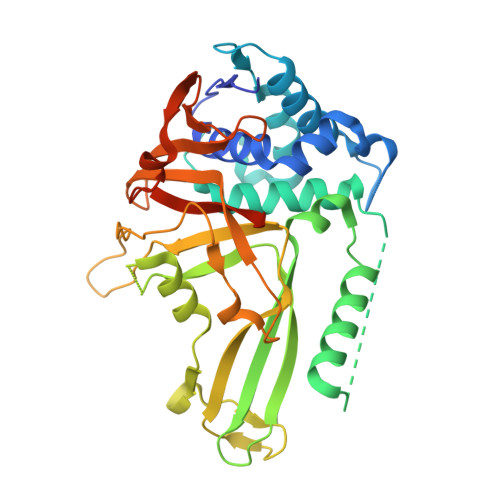
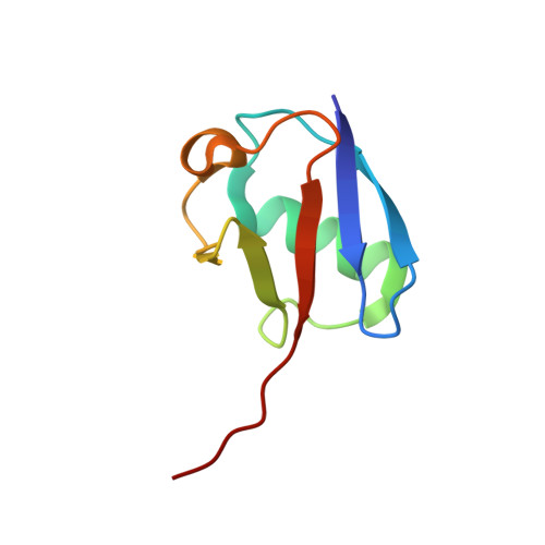
Structural Basis of Ubiquitin Recognition by the Deubiquitinating Protease USP2.

Renatus, M.Parrado, S.G.D'Arcy, A.Eidhoff, U.Gerhartz, B.Hassiepen, U.Pierrat, B.Riedl, R.Vinzenz, D.Worpenberg, S.Kroemer, M.

(2006) Structure 14: 1293-1302

  • DOI: https://doi.org/10.1016/j.str.2006.06.012
  • Primary Citation of Related Structures:  
    2HD5

  • PubMed Abstract: 

    Deubiquitinating proteases reverse protein ubiquitination and rescue their target proteins from destruction by the proteasome. USP2, a cysteine protease and a member of the ubiquitin specific protease family, is overexpressed in prostate cancer and stabilizes fatty acid synthase, which has been associated with the malignancy of some aggressive prostate cancers. Here, we report the structure of the human USP2 catalytic domain in complex with ubiquitin. Ubiquitin uses two major sites for the interaction with the protease. Both sites are required simultaneously, as shown by USP2 inhibition assays with peptides and ubiquitin mutants. In addition, a layer of ordered water molecules mediates key interactions between ubiquitin and USP2. As several of those molecules are found at identical positions in the previously solved USP7/ubiquitin-aldehyde complex structure, we suggest a general mechanism of water-mediated ubiquitin recognition by USPs.


  • Organizational Affiliation

    Protease Platform, Novartis Institutes for BioMedical Research, 4002 Basel, Switzerland. martin.renatus@novartis.com


Macromolecules
Find similar proteins by:  (by identity cutoff)  |  3D Structure
Entity ID: 1
MoleculeChains Sequence LengthOrganismDetailsImage
Ubiquitin carboxyl-terminal hydrolase 2359Homo sapiensMutation(s): 0 
Gene Names: USP2UBP41
EC: 3.1.2.15
UniProt & NIH Common Fund Data Resources
Find proteins for O75604 (Homo sapiens)
Explore O75604 
Go to UniProtKB:  O75604
PHAROS:  O75604
GTEx:  ENSG00000036672 
Entity Groups  
Sequence Clusters30% Identity50% Identity70% Identity90% Identity95% Identity100% Identity
UniProt GroupO75604
Sequence Annotations
Expand
  • Reference Sequence
Find similar proteins by:  (by identity cutoff)  |  3D Structure
Entity ID: 2
MoleculeChains Sequence LengthOrganismDetailsImage
Polyubiquitin76Bos taurusMutation(s): 0 
UniProt
Find proteins for P0CH28 (Bos taurus)
Explore P0CH28 
Go to UniProtKB:  P0CH28
Entity Groups  
Sequence Clusters30% Identity50% Identity70% Identity90% Identity95% Identity100% Identity
UniProt GroupP0CH28
Sequence Annotations
Expand
  • Reference Sequence
Small Molecules
Ligands 1 Unique
IDChains Name / Formula / InChI Key2D Diagram3D Interactions
ZN
Query on ZN

Download Ideal Coordinates CCD File 
C [auth A]ZINC ION
Zn
PTFCDOFLOPIGGS-UHFFFAOYSA-N
Experimental Data & Validation

Experimental Data

  • Method: X-RAY DIFFRACTION
  • Resolution: 1.85 Å
  • R-Value Free: 0.229 
  • R-Value Work: 0.177 
  • Space Group: C 1 2 1
Unit Cell:
Length ( Å )Angle ( ˚ )
a = 106.903α = 90
b = 45.534β = 110.01
c = 76.36γ = 90
Software Package:
Software NamePurpose
DENZOdata reduction
SCALEPACKdata scaling
PHASERphasing
CNSrefinement
PDB_EXTRACTdata extraction
CrystalCleardata reduction
CNXrefinement

Structure Validation

View Full Validation Report


Entry History 

Deposition Data

Revision History  (Full details and data files)

  • Version 1.0: 2006-08-15
    Type: Initial release
  • Version 1.1: 2008-05-01
    Changes: Version format compliance
  • Version 1.2: 2011-07-13
    Changes: Version format compliance
  • Version 1.3: 2017-10-18
    Changes: Refinement description
  • Version 1.4: 2023-08-30
    Changes: Data collection, Database references, Derived calculations, Refinement description