CgNa, a type I toxin from the giant Caribbean sea anemone Condylactis gigantea shows structural similarities to both type I and II toxins, as well as distinctive structural and functional properties(1).
Salceda, E., Perez-Castells, J., Lopez-Mendez, B., Garateix, A., Salazar, H., Lopez, O., Aneiros, A., Standker, L., Beress, L., Forssmann, W.G., Soto, E., Jimenez-Barbero, J., Gimenez-Gallego, G.(2007) Biochem J 406: 67-76
- PubMed: 17506725
- DOI: https://doi.org/10.1042/BJ20070130
- Primary Citation of Related Structures:
2H9X - PubMed Abstract:
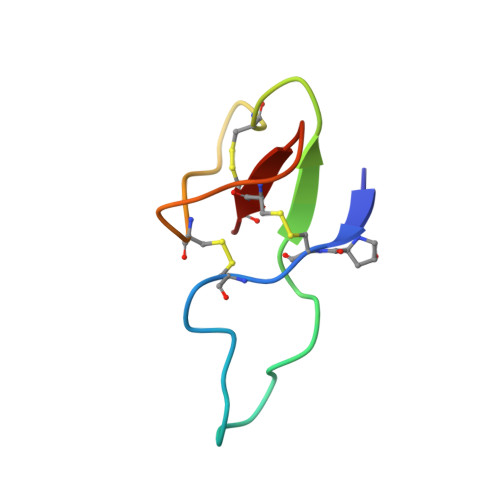
CgNa (Condylactis gigantea neurotoxin) is a 47-amino-acid- residue toxin from the giant Caribbean sea anemone Condylactis gigantea. The structure of CgNa, which was solved by 1H-NMR spectroscopy, is somewhat atypical and displays significant homology with both type I and II anemone toxins. CgNa also displays a considerable number of exceptions to the canonical structural elements that are thought to be essential for the activity of this group of toxins. Furthermore, unique residues in CgNa define a characteristic structure with strong negatively charged surface patches. These patches disrupt a surface-exposed cluster of hydrophobic residues present in all anemone-derived toxins described to date. A thorough characterization by patch-clamp analysis using rat DRG (dorsal root ganglion) neurons indicated that CgNa preferentially binds to TTX-S (tetrodotoxin-sensitive) voltage-gated sodium channels in the resting state. This association increased the inactivation time constant and the rate of recovery from inactivation, inducing a significant shift in the steady state of inactivation curve to the left. The specific structural features of CgNa may explain its weaker inhibitory capacity when compared with the other type I and II anemone toxins.
Organizational Affiliation:
Instituto de Fisiología, Universidad Autónoma de Puebla, 14 Sur 6301, 72570 Puebla, México.