2H6J

Crystal Structure of the Beta F145A Rhodococcus Proteasome


Experimental Data Snapshot

  • Method: X-RAY DIFFRACTION
  • Resolution: 3.20 Å
  • R-Value Free: 0.290 
  • R-Value Work: 0.262 
  • R-Value Observed: 0.262 

wwPDB Validation   3D Report Full Report


This is version 1.5 of the entry. See complete history


Literature

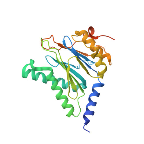
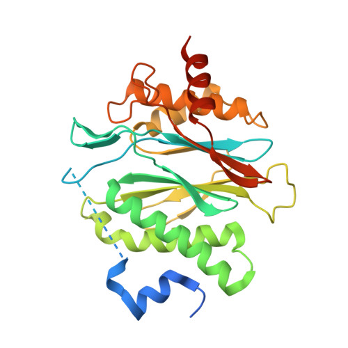
Proteasome assembly triggers a switch required for active-site maturation.

Witt, S.Kwon, Y.D.Sharon, M.Felderer, K.Beuttler, M.Robinson, C.V.Baumeister, W.Jap, B.K.

(2006) Structure 14: 1179-1188

  • DOI: https://doi.org/10.1016/j.str.2006.05.019
  • Primary Citation of Related Structures:  
    2H6J

  • PubMed Abstract: 

    The processing of propeptides and the maturation of 20S proteasomes require the association of beta rings from two half proteasomes. We propose an assembly-dependent activation model in which interactions between helix (H3 and H4) residues of the opposing half proteasomes are prerequisite for appropriate positioning of the S2-S3 loop; such positioning enables correct coordination of the active-site residue needed for propeptide cleavage. Mutations of H3 or H4 residues that participate in the association of two half proteasomes inhibit activation and prevent, in nearly all cases, the formation of full proteasomes. In contrast, mutations affecting interactions with residues of the S2-S3 loop allow the assembly of full, but activity impacted, proteasomes. The crystal structure of the inactive H3 mutant, Phe145Ala, shows that the S2-S3 loop is displaced from the position observed in wild-type proteasomes. These data support the proposed assembly-dependent activation model in which the S2-S3 loop acts as an activation switch.


  • Organizational Affiliation

    Department of Molecular Structural Biology, Max-Planck-Institute of Biochemistry, Martinsried 82152, Germany.


Macromolecules
Find similar proteins by:  (by identity cutoff)  |  3D Structure
Entity ID: 1
MoleculeChains Sequence LengthOrganismDetailsImage
Proteasome alpha-type subunit 1
A, B, C, D, E
A, B, C, D, E, F, G
259Rhodococcus erythropolisMutation(s): 0 
Gene Names: prcA 1
EC: 3.4.25.1
UniProt
Find proteins for Q53080 (Rhodococcus erythropolis)
Explore Q53080 
Go to UniProtKB:  Q53080
Entity Groups  
Sequence Clusters30% Identity50% Identity70% Identity90% Identity95% Identity100% Identity
UniProt GroupQ53080
Sequence Annotations
Expand
  • Reference Sequence
Find similar proteins by:  (by identity cutoff)  |  3D Structure
Entity ID: 2
MoleculeChains Sequence LengthOrganismDetailsImage
Proteasome beta-type subunit 1
H, I, J, K, L
H, I, J, K, L, M, N
294Rhodococcus erythropolisMutation(s): 1 
Gene Names: prcB 1
EC: 3.4.25.1
UniProt
Find proteins for Q53079 (Rhodococcus erythropolis)
Explore Q53079 
Go to UniProtKB:  Q53079
Entity Groups  
Sequence Clusters30% Identity50% Identity70% Identity90% Identity95% Identity100% Identity
UniProt GroupQ53079
Sequence Annotations
Expand
  • Reference Sequence
Experimental Data & Validation

Experimental Data

  • Method: X-RAY DIFFRACTION
  • Resolution: 3.20 Å
  • R-Value Free: 0.290 
  • R-Value Work: 0.262 
  • R-Value Observed: 0.262 
  • Space Group: C 2 2 21
Unit Cell:
Length ( Å )Angle ( ˚ )
a = 149.437α = 90
b = 210.714β = 90
c = 248.126γ = 90
Software Package:
Software NamePurpose
DENZOdata reduction
SCALEPACKdata scaling
CNSrefinement
PDB_EXTRACTdata extraction
ADSCdata collection
CNSphasing

Structure Validation

View Full Validation Report


Entry History 

Deposition Data

  • Released Date: 2006-08-01 
  • Deposition Author(s): Kwon, Y.D.

Revision History  (Full details and data files)

  • Version 1.0: 2006-08-01
    Type: Initial release
  • Version 1.1: 2008-05-01
    Changes: Version format compliance
  • Version 1.2: 2011-07-13
    Changes: Derived calculations, Version format compliance
  • Version 1.3: 2017-10-18
    Changes: Refinement description, Structure summary
  • Version 1.4: 2021-10-20
    Changes: Database references
  • Version 1.5: 2023-08-30
    Changes: Data collection, Refinement description