2H65

Crystal strusture of caspase-3 with inhibitor Ac-VDVAD-Cho


Experimental Data Snapshot

  • Method: X-RAY DIFFRACTION
  • Resolution: 2.30 Å
  • R-Value Free: 0.246 
  • R-Value Work: 0.209 
  • R-Value Observed: 0.198 

wwPDB Validation   3D Report Full Report


This is version 1.5 of the entry. See complete history


Literature

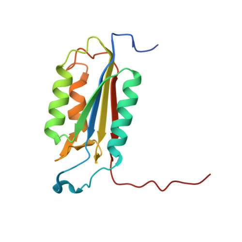
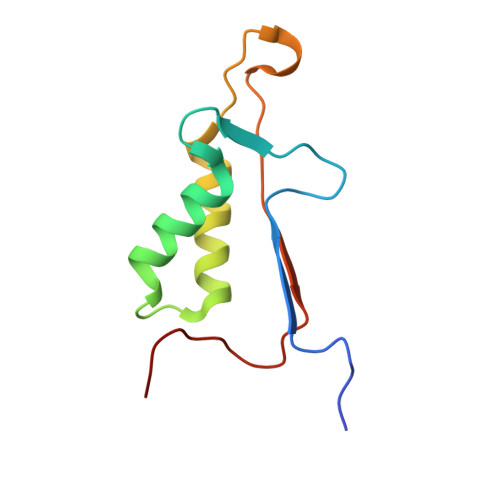
Structural and kinetic analysis of caspase-3 reveals role for s5 binding site in substrate recognition

Fang, B.Boross, P.I.Tozser, J.Weber, I.T.

(2006) J Mol Biol 360: 654-666

  • DOI: https://doi.org/10.1016/j.jmb.2006.05.041
  • Primary Citation of Related Structures:  
    2H5I, 2H5J, 2H65

  • PubMed Abstract: 

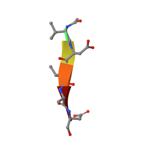
    The molecular basis for the substrate specificity of human caspase-3 has been investigated using peptide analog inhibitors and substrates that vary at the P2, P3, and P5 positions. Crystal structures were determined of caspase-3 complexes with the substrate analogs at resolutions of 1.7 A to 2.3 A. Differences in the interactions of caspase-3 with the analogs are consistent with the Ki values of 1.3 nM, 6.5 nM, and 12.4 nM for Ac-DEVD-Cho, Ac-VDVAD-Cho and Ac-DMQD-Cho, respectively, and relative kcat/Km values of 100%, 37% and 17% for the corresponding peptide substrates. The bound peptide analogs show very similar interactions for the main-chain atoms and the conserved P1 Asp and P4 Asp, while interactions vary for P2 and P3. P2 lies in a hydrophobic S2 groove, consistent with the weaker inhibition of Ac-DMQD-Cho with polar P2 Gln. S3 is a surface hydrophilic site with favorable polar interactions with P3 Glu in Ac-DEVD-Cho. Ac-DMQD-Cho and Ac-VDVAD-Cho have hydrophobic P3 residues that are not optimal in the polar S3 site, consistent with their weaker inhibition. A hydrophobic S5 site was identified for caspase-3, where the side-chains of Phe250 and Phe252 interact with P5 Val of Ac-VDVAD-Cho, and enclose the substrate-binding site by conformational change. The kinetic importance of hydrophobic P5 residues was confirmed by more efficient hydrolysis of caspase-3 substrates Ac-VDVAD-pNA and Ac-LDVAD-pNA compared with Ac-DVAD-pNA. In contrast, caspase-7 showed less efficient hydrolysis of the substrates with P5 Val or Leu compared with Ac-DVAD-pNA. Caspase-3 and caspase-2 share similar hydrophobic S5 sites, while caspases 1, 7, 8 and 9 do not have structurally equivalent hydrophobic residues; these caspases are likely to differ in their selectivity for the P5 position of substrates. The distinct selectivity for P5 will help define the particular substrates and signaling pathways associated with each caspase.


  • Organizational Affiliation

    Department of Biology, Molecular Basis of Disease, Georgia State University, Atlanta, GA 30303, USA.


Macromolecules
Find similar proteins by:  (by identity cutoff)  |  3D Structure
Entity ID: 1
MoleculeChains Sequence LengthOrganismDetailsImage
caspase-3, p17 subunit
A, C
146Homo sapiensMutation(s): 0 
Gene Names: CASP3CPP32
EC: 3.4.22
UniProt & NIH Common Fund Data Resources
Find proteins for P42574 (Homo sapiens)
Explore P42574 
Go to UniProtKB:  P42574
PHAROS:  P42574
GTEx:  ENSG00000164305 
Entity Groups  
Sequence Clusters30% Identity50% Identity70% Identity90% Identity95% Identity100% Identity
UniProt GroupP42574
Sequence Annotations
Expand
  • Reference Sequence
Find similar proteins by:  (by identity cutoff)  |  3D Structure
Entity ID: 2
MoleculeChains Sequence LengthOrganismDetailsImage
caspase-3, p12 subunit
B, D
95Homo sapiensMutation(s): 0 
Gene Names: CASP3CPP32
EC: 3.4.22
UniProt & NIH Common Fund Data Resources
Find proteins for P42574 (Homo sapiens)
Explore P42574 
Go to UniProtKB:  P42574
PHAROS:  P42574
GTEx:  ENSG00000164305 
Entity Groups  
Sequence Clusters30% Identity50% Identity70% Identity90% Identity95% Identity100% Identity
UniProt GroupP42574
Sequence Annotations
Expand
  • Reference Sequence

Find similar proteins by:  Sequence   |   3D Structure  

Entity ID: 3
MoleculeChains Sequence LengthOrganismDetailsImage
Ac-VDVAD-Cho
E, F
6N/AMutation(s): 0 
Sequence Annotations
Expand
  • Reference Sequence
Biologically Interesting Molecules (External Reference) 1 Unique
Experimental Data & Validation

Experimental Data

  • Method: X-RAY DIFFRACTION
  • Resolution: 2.30 Å
  • R-Value Free: 0.246 
  • R-Value Work: 0.209 
  • R-Value Observed: 0.198 
  • Space Group: P 1 21 1
Unit Cell:
Length ( Å )Angle ( ˚ )
a = 49.89α = 90
b = 67.4β = 100.8
c = 93.49γ = 90
Software Package:
Software NamePurpose
MADCCDdata collection
HKL-2000data reduction
AMoREphasing
CNSrefinement
MADCCDdata reduction
HKL-2000data scaling

Structure Validation

View Full Validation Report


Entry History 

Deposition Data

Revision History  (Full details and data files)

  • Version 1.0: 2006-09-19
    Type: Initial release
  • Version 1.1: 2008-05-01
    Changes: Version format compliance
  • Version 1.2: 2011-07-13
    Changes: Atomic model, Database references, Derived calculations, Non-polymer description, Structure summary, Version format compliance
  • Version 1.3: 2012-12-12
    Changes: Other
  • Version 1.4: 2023-08-30
    Changes: Data collection, Database references, Derived calculations, Refinement description
  • Version 1.5: 2023-11-15
    Changes: Data collection