


Crystal structure of hypothetical protein PG_1108 from Porphyromonas gingivalis W83
Nocek, B., Bigelow, L., Moy, S., Joachimiak, A.To be published.
Experimental Data Snapshot
wwPDB Validation 3D Report Full Report

Entity ID: 1 | |||||
|---|---|---|---|---|---|
| Molecule | Chains | Sequence Length | Organism | Details | Image |
| Hypothetical protein PG_1108 | 133 | Porphyromonas gingivalis | Mutation(s): 0 | 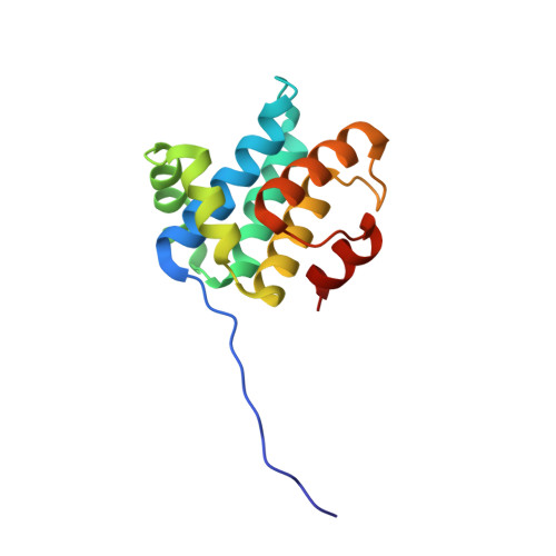 | |
UniProt | |||||
Find proteins for Q7MVF6 (Porphyromonas gingivalis (strain ATCC BAA-308 / W83)) Explore Q7MVF6 Go to UniProtKB: Q7MVF6 | |||||
Entity Groups | |||||
| Sequence Clusters | 30% Identity50% Identity70% Identity90% Identity95% Identity100% Identity | ||||
| UniProt Group | Q7MVF6 | ||||
Sequence AnnotationsExpand | |||||
| |||||
| Ligands 1 Unique | |||||
|---|---|---|---|---|---|
| ID | Chains | Name / Formula / InChI Key | 2D Diagram | 3D Interactions | |
| MG Query on MG | E [auth A], F [auth B], G [auth C] | MAGNESIUM ION Mg JLVVSXFLKOJNIY-UHFFFAOYSA-N |  | ||
| Length ( Å ) | Angle ( ˚ ) |
|---|---|
| a = 79.961 | α = 90 |
| b = 84.826 | β = 90 |
| c = 164.535 | γ = 90 |
| Software Name | Purpose |
|---|---|
| REFMAC | refinement |
| SBC-Collect | data collection |
| HKL-2000 | data scaling |
| HKL-3000 | phasing |
| SHELXD | phasing |
| SHELXE | model building |
| MLPHARE | phasing |
| DM | phasing |
| SOLVE | phasing |
| RESOLVE | phasing |
| Coot | model building |
| CCP4 | phasing |
RCSB PDB (citation) is hosted by
RCSB PDB is a member of the

