2H5D

0.9A resolution crystal structure of alpha-lytic protease complexed with a transition state analogue, MeOSuc-Ala-Ala-Pro-Val boronic acid


Experimental Data Snapshot

  • Method: X-RAY DIFFRACTION
  • Resolution: 0.90 Å
  • R-Value Free: 0.091 
  • R-Value Observed: 0.080 

wwPDB Validation   3D Report Full Report


This is version 2.0 of the entry. See complete history


Literature

Subangstrom crystallography reveals that short ionic hydrogen bonds, and not a His-Asp low-barrier hydrogen bond, stabilize the transition state in serine protease catalysis

Fuhrmann, C.N.Daugherty, M.D.Agard, D.A.

(2006) J Am Chem Soc 128: 9086-9102

  • DOI: https://doi.org/10.1021/ja057721o

  • PubMed Abstract: 

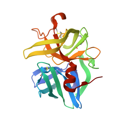
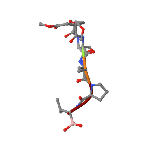
    To address questions regarding the mechanism of serine protease catalysis, we have solved two X-ray crystal structures of alpha-lytic protease (alphaLP) that mimic aspects of the transition states: alphaLP at pH 5 (0.82 A resolution) and alphaLP bound to the peptidyl boronic acid inhibitor, MeOSuc-Ala-Ala-Pro-boroVal (0.90 A resolution). Based on these structures, there is no evidence of, or requirement for, histidine-flipping during the acylation step of the reaction. Rather, our data suggests that upon protonation of His57, Ser195 undergoes a conformational change that destabilizes the His57-Ser195 hydrogen bond, preventing the back-reaction. In both structures the His57-Asp102 hydrogen bond in the catalytic triad is a normal ionic hydrogen bond, and not a low-barrier hydrogen bond (LBHB) as previously hypothesized. We propose that the enzyme has evolved a network of relatively short hydrogen bonds that collectively stabilize the transition states. In particular, a short ionic hydrogen bond (SIHB) between His57 Nepsilon2 and the substrate's leaving group may promote forward progression of the TI1-to-acylenzyme reaction. We provide experimental evidence that refutes use of either a short donor-acceptor distance or a downfield 1H chemical shift as sole indicators of a LBHB.


  • Organizational Affiliation

    Howard Hughes Medical Institute and Department of Biochemistry and Biophysics, University of California, San Francisco, San Francisco, California 94143-2240, USA.


Macromolecules
Find similar proteins by:  (by identity cutoff)  |  3D Structure
Entity ID: 1
MoleculeChains Sequence LengthOrganismDetailsImage
ALPHA-LYTIC PROTEASE198Lysobacter enzymogenesMutation(s): 0 
EC: 3.4.21.12
UniProt
Find proteins for P00778 (Lysobacter enzymogenes)
Explore P00778 
Go to UniProtKB:  P00778
Entity Groups  
Sequence Clusters30% Identity50% Identity70% Identity90% Identity95% Identity100% Identity
UniProt GroupP00778
Sequence Annotations
Expand
  • Reference Sequence

Find similar proteins by:  Sequence   |   3D Structure  

Entity ID: 2
MoleculeChains Sequence LengthOrganismDetailsImage
MEOSUC-ALA-ALA-PRO-ALA BORONIC ACID INHIBITOR5N/AMutation(s): 0 
Sequence Annotations
Expand
  • Reference Sequence
Small Molecules
Modified Residues  1 Unique
IDChains TypeFormula2D DiagramParent
B2V
Query on B2V
B
PEPTIDE-LIKEC4 H12 B N O2VAL
Biologically Interesting Molecules (External Reference) 1 Unique
Experimental Data & Validation

Experimental Data

  • Method: X-RAY DIFFRACTION
  • Resolution: 0.90 Å
  • R-Value Free: 0.091 
  • R-Value Observed: 0.080 
  • Space Group: P 32 2 1
Unit Cell:
Length ( Å )Angle ( ˚ )
a = 65.768α = 90
b = 65.768β = 90
c = 79.554γ = 120
Software Package:
Software NamePurpose
HKL-2000data collection
SCALEPACKdata scaling
startingmodel building
SHELXL-97refinement
HKL-2000data reduction

Structure Validation

View Full Validation Report


Entry History 

Deposition Data

Revision History  (Full details and data files)

  • Version 1.0: 2006-09-26
    Type: Initial release
  • Version 1.1: 2008-05-01
    Changes: Version format compliance
  • Version 1.2: 2011-07-13
    Changes: Atomic model, Database references, Derived calculations, Non-polymer description, Structure summary, Version format compliance
  • Version 1.3: 2012-12-12
    Changes: Other
  • Version 1.4: 2023-08-30
    Changes: Data collection, Database references, Derived calculations, Refinement description
  • Version 2.0: 2023-11-15
    Changes: Atomic model, Data collection