2H1W

Crystal structure of the His183Ala mutant variant of Bacillus subtilis ferrochelatase


Experimental Data Snapshot

  • Method: X-RAY DIFFRACTION
  • Resolution: 2.60 Å
  • R-Value Free: 0.259 
  • R-Value Work: 0.218 
  • R-Value Observed: 0.218 

wwPDB Validation   3D Report Full Report


This is version 1.6 of the entry. See complete history


Literature

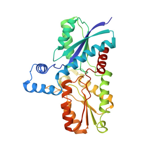
Amino Acid Residues His183 and Glu264 in Bacillus subtilis Ferrochelatase Direct and Facilitate the Insertion of Metal Ion into Protoporphyrin IX

Hansson, M.D.Karlberg, T.Rahardja, M.A.Al-Karadaghi, S.Hansson, M.

(2007) Biochemistry 46: 87-94

  • DOI: https://doi.org/10.1021/bi061760a
  • Primary Citation of Related Structures:  
    2H1V, 2H1W, 2HK6

  • PubMed Abstract: 

    Ferrochelatase catalyzes the terminal step in the heme biosynthetic pathway, i.e., the incorporation of Fe(II) into protoporphyrin IX. Various biochemical and biophysical methods have been used to probe the enzyme for metal binding residues and the location of the active site. However, the location of the metal binding site and the path of the metal into the porphyrin are still disputed. Using site-directed mutagenesis on Bacillus subtilis ferrochelatase we demonstrate that exchange of the conserved residues His183 and Glu264 affects the metal affinity of the enzyme. We also present the first X-ray crystal structure of ferrochelatase with iron. Only a single iron was found in the active site, coordinated in a square pyramidal fashion by two amino acid residues, His183 and Glu264, and three water molecules. This iron was not present in the structure of a His183Ala modified ferrochelatase. The results strongly suggest that the insertion of a metal ion into protoporphyrin IX by ferrochelatase occurs from a metal binding site represented by His183 and Glu264.


  • Organizational Affiliation

    Department of Biochemistry and Department of Molecular Biophysics, Lund University, Box 124, 221 00 Lund, Sweden. matthias.hansson@biochemistry.lu.se


Macromolecules
Find similar proteins by:  (by identity cutoff)  |  3D Structure
Entity ID: 1
MoleculeChains Sequence LengthOrganismDetailsImage
Ferrochelatase310Bacillus subtilisMutation(s): 1 
Gene Names: hemHhemF
EC: 4.99.1.1
UniProt
Find proteins for P32396 (Bacillus subtilis (strain 168))
Explore P32396 
Go to UniProtKB:  P32396
Entity Groups  
Sequence Clusters30% Identity50% Identity70% Identity90% Identity95% Identity100% Identity
UniProt GroupP32396
Sequence Annotations
Expand
  • Reference Sequence
Experimental Data & Validation

Experimental Data

  • Method: X-RAY DIFFRACTION
  • Resolution: 2.60 Å
  • R-Value Free: 0.259 
  • R-Value Work: 0.218 
  • R-Value Observed: 0.218 
  • Space Group: P 21 21 21
Unit Cell:
Length ( Å )Angle ( ˚ )
a = 47.94α = 90
b = 49.72β = 90
c = 116.54γ = 90
Software Package:
Software NamePurpose
CNSrefinement
XDSdata reduction
XDSdata scaling
CNSphasing

Structure Validation

View Full Validation Report


Entry History 

Deposition Data

Revision History  (Full details and data files)

  • Version 1.0: 2007-01-16
    Type: Initial release
  • Version 1.1: 2008-05-01
    Changes: Version format compliance
  • Version 1.2: 2011-07-13
    Changes: Version format compliance
  • Version 1.3: 2014-11-12
    Changes: Structure summary
  • Version 1.4: 2018-03-07
    Changes: Data collection
  • Version 1.5: 2021-10-20
    Changes: Database references, Derived calculations
  • Version 1.6: 2023-08-30
    Changes: Data collection, Refinement description