2GYV

Crystal structure of Mus musculus Acetylcholinesterase in complex with Ortho-7


Experimental Data Snapshot

  • Method: X-RAY DIFFRACTION
  • Resolution: 2.50 Å
  • R-Value Free: 0.241 
  • R-Value Work: 0.197 
  • R-Value Observed: 0.198 

wwPDB Validation   3D Report Full Report


Ligand Structure Quality Assessment 


This is version 1.5 of the entry. See complete history


Literature

Crystal structures of acetylcholinesterase in complex with HI-6, Ortho-7 and obidoxime: Structural basis for differences in the ability to reactivate tabun conjugates.

Ekstrom, F.Pang, Y.P.Boman, M.Artursson, E.Akfur, C.Borjegren, S.

(2006) Biochem Pharmacol 72: 597-607

  • DOI: https://doi.org/10.1016/j.bcp.2006.05.027
  • Primary Citation of Related Structures:  
    2GYU, 2GYV, 2GYW

  • PubMed Abstract: 

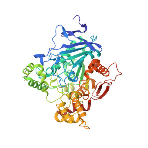
    Inhibition of acetylcholinesterase (AChE) by organophosphorus compounds (OPs) such as pesticides and nerve agents causes acute toxicity or death of the intoxicated individual. The inhibited AChE may be reactivated by certain oximes as antidotes for clinical treatment of OP-intoxications. Crystal structures of the oximes HI-6, Ortho-7 and obidoxime in complex with Mus musculus acetylcholinesterase (mAChE) reveal different roles of the peripheral anionic site (PAS) in the binding of the oximes. A limited structural change of the side chains of Trp286 and Asp74 facilitates the intercalation of the 4-carboxylamide pyridinium ring of HI-6 between the side chains of Tyr124 and Trp286. The 2-carboxyimino pyridinium ring of HI-6 is accommodated at the entrance of the catalytic site with the oximate forming a hydrogen bond to the main-chain nitrogen atom of Phe295. In contrast to HI-6, the coordination of Ortho-7 and obidoxime within the PAS is facilitated by an extended structural change of Trp286 that allows one of the carboxyimino pyridinium rings to form a cation-pi interaction with the aromatic groups of Tyr72 and Trp286. The central chain of Ortho-7 and obidoxime is loosely coordinated in the active-site gorge, whereas the second carboxyimino pyridinium ring is accommodated in the vicinity of the phenol ring of Tyr337. The structural data clearly show analogous coordination of Ortho-7 and obidoxime within the active-site gorge of AChE. Different ability to reactivate AChE inhibited by tabun is shown in end-point reactivation experiments where HI-6, Ortho-7 and obidoxime showed an efficiency of 1, 45 and 38%, respectively. The low efficiency of HI-6 and the significantly higher efficiency of Ortho-7 and obidoxime may be explained by the differential binding of the oximes in the PAS and active-site gorge of AChE.


  • Organizational Affiliation

    Swedish Defense Research Agency, Division of NBC Defense, S-901 82, Umeå, Sweden. fredrik.ekstrom@foi.se


Macromolecules
Find similar proteins by:  (by identity cutoff)  |  3D Structure
Entity ID: 1
MoleculeChains Sequence LengthOrganismDetailsImage
Acetylcholinesterase
A, B
543Mus musculusMutation(s): 0 
Gene Names: Ache
EC: 3.1.1.7
UniProt & NIH Common Fund Data Resources
Find proteins for P21836 (Mus musculus)
Explore P21836 
Go to UniProtKB:  P21836
IMPC:  MGI:87876
Entity Groups  
Sequence Clusters30% Identity50% Identity70% Identity90% Identity95% Identity100% Identity
UniProt GroupP21836
Sequence Annotations
Expand
  • Reference Sequence
Small Molecules
Ligands 4 Unique
IDChains Name / Formula / InChI Key2D Diagram3D Interactions
HBP
Query on HBP

Download Ideal Coordinates CCD File 
F [auth A],
H [auth B]
1,7-HEPTYLENE-BIS-N,N'-SYN-2-PYRIDINIUMALDOXIME
C19 H26 N4 O2
NJSMUQMHXZRXQC-UHFFFAOYSA-P
P3G
Query on P3G

Download Ideal Coordinates CCD File 
I [auth B]3,6,9,12,15-PENTAOXAHEPTADECANE
C12 H26 O5
HYDWALOBQJFOMS-UHFFFAOYSA-N
NAG
Query on NAG

Download Ideal Coordinates CCD File 
C [auth A],
D [auth A]
2-acetamido-2-deoxy-beta-D-glucopyranose
C8 H15 N O6
OVRNDRQMDRJTHS-FMDGEEDCSA-N
CO3
Query on CO3

Download Ideal Coordinates CCD File 
E [auth A],
G [auth B]
CARBONATE ION
C O3
BVKZGUZCCUSVTD-UHFFFAOYSA-L
Experimental Data & Validation

Experimental Data

  • Method: X-RAY DIFFRACTION
  • Resolution: 2.50 Å
  • R-Value Free: 0.241 
  • R-Value Work: 0.197 
  • R-Value Observed: 0.198 
  • Space Group: P 21 21 21
Unit Cell:
Length ( Å )Angle ( ˚ )
a = 76.73α = 90
b = 108.58β = 90
c = 220.58γ = 90
Software Package:
Software NamePurpose
REFMACrefinement
PDB_EXTRACTdata extraction
XDSdata scaling

Structure Validation

View Full Validation Report


Ligand Structure Quality Assessment 


Entry History 

Deposition Data

Revision History  (Full details and data files)

  • Version 1.0: 2006-08-15
    Type: Initial release
  • Version 1.1: 2008-05-01
    Changes: Version format compliance
  • Version 1.2: 2011-07-13
    Changes: Advisory, Version format compliance
  • Version 1.3: 2018-03-07
    Changes: Data collection
  • Version 1.4: 2020-07-29
    Type: Remediation
    Reason: Carbohydrate remediation
    Changes: Data collection, Derived calculations, Structure summary
  • Version 1.5: 2023-08-30
    Changes: Data collection, Database references, Refinement description, Structure summary