2GWW

Human vinculin (head domain, Vh1, residues 1-258) in complex with Shigella's IpaA vinculin binding site (residues 602-633)


Experimental Data Snapshot

  • Method: X-RAY DIFFRACTION
  • Resolution: 2.72 Å
  • R-Value Free: 0.291 
  • R-Value Work: 0.229 
  • R-Value Observed: 0.232 

wwPDB Validation   3D Report Full Report


This is version 1.3 of the entry. See complete history


Literature

Shigella applies molecular mimicry to subvert vinculin and invade host cells.

Izard, T.Tran Van Nhieu, G.Bois, P.R.

(2006) J Cell Biol 175: 465-475

  • DOI: https://doi.org/10.1083/jcb.200605091
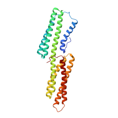
  • Primary Citation of Related Structures:  
    2GWW, 2HSQ

  • PubMed Abstract: 

    Shigella flexneri, the causative agent of bacillary dysentery, injects invasin proteins through a type III secretion apparatus upon contacting the host cell, which triggers pathogen internalization. The invasin IpaA is essential for S. flexneri pathogenesis and binds to the cytoskeletal protein vinculin to facilitate host cell entry. We report that IpaA harbors two vinculin-binding sites (VBSs) within its C-terminal domain that bind to and activate vinculin in a mutually exclusive fashion. Only the highest affinity C-terminal IpaA VBS is necessary for efficient entry and cell-cell spread of S. flexneri, whereas the lower affinity VBS appears to contribute to vinculin recruitment at entry foci of the pathogen. Finally, the crystal structures of vinculin in complex with the VBSs of IpaA reveal the mechanism by which IpaA subverts vinculin's functions, where S. flexneri utilizes a remarkable level of molecular mimicry of the talin-vinculin interaction to activate vinculin. Mimicry of vinculin's interactions may therefore be a general mechanism applied by pathogens to infect the host cell.


  • Organizational Affiliation

    Cell Adhesion Laboratory, Department of Oncology, St. Jude Children's Research Hospital, Memphis, TN 38105, USA. tina.izard@stjude.org


Macromolecules
Find similar proteins by:  (by identity cutoff)  |  3D Structure
Entity ID: 1
MoleculeChains Sequence LengthOrganismDetailsImage
vinculin266Homo sapiensMutation(s): 0 
UniProt & NIH Common Fund Data Resources
Find proteins for P18206 (Homo sapiens)
Explore P18206 
Go to UniProtKB:  P18206
PHAROS:  P18206
GTEx:  ENSG00000035403 
Entity Groups  
Sequence Clusters30% Identity50% Identity70% Identity90% Identity95% Identity100% Identity
UniProt GroupP18206
Sequence Annotations
Expand
  • Reference Sequence
Find similar proteins by:  (by identity cutoff)  |  3D Structure
Entity ID: 2
MoleculeChains Sequence LengthOrganismDetailsImage
IpaA30Shigella flexneriMutation(s): 0 
UniProt
Find proteins for P18010 (Shigella flexneri)
Explore P18010 
Go to UniProtKB:  P18010
Entity Groups  
Sequence Clusters30% Identity50% Identity70% Identity90% Identity95% Identity100% Identity
UniProt GroupP18010
Sequence Annotations
Expand
  • Reference Sequence
Experimental Data & Validation

Experimental Data

  • Method: X-RAY DIFFRACTION
  • Resolution: 2.72 Å
  • R-Value Free: 0.291 
  • R-Value Work: 0.229 
  • R-Value Observed: 0.232 
  • Space Group: I 41 3 2
Unit Cell:
Length ( Å )Angle ( ˚ )
a = 151.31α = 90
b = 151.31β = 90
c = 151.31γ = 90
Software Package:
Software NamePurpose
BUSTER-TNTrefinement

Structure Validation

View Full Validation Report


Entry History 

Deposition Data

  • Released Date: 2006-11-14 
  • Deposition Author(s): Izard, T.

Revision History  (Full details and data files)

  • Version 1.0: 2006-11-14
    Type: Initial release
  • Version 1.1: 2008-05-01
    Changes: Version format compliance
  • Version 1.2: 2011-07-13
    Changes: Version format compliance
  • Version 1.3: 2024-02-14
    Changes: Data collection, Database references