Domain orientation in the inactive response regulator Mycobacterium tuberculosis MtrA provides a barrier to activation.
Friedland, N., Mack, T.R., Yu, M., Hung, L.W., Terwilliger, T.C., Waldo, G.S., Stock, A.M.(2007) Biochemistry 46: 6733-6743
- PubMed: 17511470
- DOI: https://doi.org/10.1021/bi602546q
- Primary Citation of Related Structures:
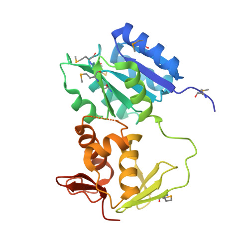
2GWR - PubMed Abstract:
The structure of MtrA, an essential gene product for the human pathogen Mycobacterium tuberculosis, has been solved to a resolution of 2.1 A. MtrA is a member of the OmpR/PhoB family of response regulators and represents the fourth family member for which a structure of the protein in its inactive state has been determined. As is true for all OmpR/PhoB family members, MtrA possesses an N-terminal regulatory domain and a C-terminal winged helix-turn-helix DNA-binding domain, with phosphorylation of the regulatory domain modulating the activity of the protein. In the inactive form of MtrA, these two domains form an extensive interface that is composed of the alpha4-beta5-alpha5 face of the regulatory domain and the C-terminal end of the positioning helix, the trans-activation loop, and the recognition helix of the DNA-binding domain. This domain orientation suggests a mechanism of mutual inhibition by the two domains. Activation of MtrA would require a disruption of this interface to allow the alpha4-beta5-alpha5 face of the regulatory domain to form the intermolecule interactions that are associated with the active state and to allow the recognition helix to interact with DNA. Furthermore, the interface appears to stabilize the inactive conformation of MtrA, potentially reducing the rate of phosphorylation of the N-terminal domain. This combination of effects may form a switch, regulating the activity of MtrA. The domain orientation exhibited by MtrA also provides a rationale for the variation in linker length that is observed within the OmpR/PhoB family of response regulators.
Organizational Affiliation:
Bioscience Division, Los Alamos National Laboratory, Los Alamos, New Mexico 87545, USA.