2GWO

crystal structure of TMDP


Experimental Data Snapshot

  • Method: X-RAY DIFFRACTION
  • Resolution: 2.40 Å
  • R-Value Free: 0.250 
  • R-Value Work: 0.198 
  • R-Value Observed: 0.198 

wwPDB Validation   3D Report Full Report


This is version 1.3 of the entry. See complete history


Literature

Crystal structure of human TMDP, a testis-specific dual specificity protein phosphatase: implications for substrate specificity

Kim, S.J.Jeong, D.G.Yoon, T.S.Son, J.H.Cho, S.K.Ryu, S.E.Kim, J.H.

(2007) Proteins 66: 239-245

  • DOI: https://doi.org/10.1002/prot.21197
  • Primary Citation of Related Structures:  
    2GWO

  • PubMed Abstract: 

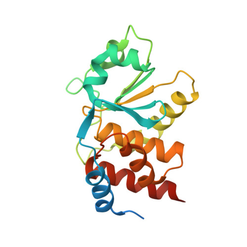
    The testis- and skeletal-muscle-specific dual-specificity phosphatase (TMDP) is a member of the dual-specificity phosphatase (DSP) subgroup of protein tyrosine phosphatases. TMDP has similar activities toward both tyrosine and threonine phosphorylated substrates, and is supposed to be involved in spermatogenesis. Here, we report the crystal structure of human TMDP at a resolution of 2.4 A. In spite of high sequence similarity with other DSPs, the crystal structure of TMDP shows distinct structural motifs and surface properties. In TMDP, the alpha1-beta1 loop, a substrate recognition motif is located further away from the active site loop in comparison to prototype DSP Vaccinia H1 related phophatase (VHR), which preferentially dephosphorylates tyrosine phosphorylated substrates and down-regulates MAP kinase signaling. Residues in the active site residues of TMDP are smaller in size and more hydrophobic than those of VHR. In addition, TMDP cannot be aligned with VHR in loop beta3-alpha4. These differences in the active site of TMDP result in a flat and wide pocket structure, allowing equal binding of phosphotyrosine and phosphothreonine substrates.


  • Organizational Affiliation

    Center for Cellular Switch Protein Structure, Korea Research Institute of Bioscience and Biotechnology, Daejeon 305-806, Republic of Korea.


Macromolecules
Find similar proteins by:  (by identity cutoff)  |  3D Structure
Entity ID: 1
MoleculeChains Sequence LengthOrganismDetailsImage
Dual specificity protein phosphatase 13
A, B, C, D
198Homo sapiensMutation(s): 0 
Gene Names: DUSP13TMDP
EC: 3.1.3.48 (PDB Primary Data), 3.1.3.16 (PDB Primary Data)
UniProt & NIH Common Fund Data Resources
Find proteins for Q9UII6 (Homo sapiens)
Explore Q9UII6 
Go to UniProtKB:  Q9UII6
PHAROS:  Q9UII6
GTEx:  ENSG00000079393 
Entity Groups  
Sequence Clusters30% Identity50% Identity70% Identity90% Identity95% Identity100% Identity
UniProt GroupQ9UII6
Sequence Annotations
Expand
  • Reference Sequence
Experimental Data & Validation

Experimental Data

  • Method: X-RAY DIFFRACTION
  • Resolution: 2.40 Å
  • R-Value Free: 0.250 
  • R-Value Work: 0.198 
  • R-Value Observed: 0.198 
  • Space Group: P 1 21 1
Unit Cell:
Length ( Å )Angle ( ˚ )
a = 61.586α = 90
b = 72.24β = 99.66
c = 89.896γ = 90
Software Package:
Software NamePurpose
MOSFLMdata reduction
SCALAdata scaling
EPMRphasing
CNSrefinement
CCP4data scaling

Structure Validation

View Full Validation Report


Entry History 

Deposition Data

Revision History  (Full details and data files)

  • Version 1.0: 2007-03-20
    Type: Initial release
  • Version 1.1: 2008-05-01
    Changes: Version format compliance
  • Version 1.2: 2011-07-13
    Changes: Version format compliance
  • Version 1.3: 2024-03-13
    Changes: Data collection, Database references