2GVE

Time-of-Flight Neutron Diffraction Structure of D-Xylose Isomerase


Experimental Data Snapshot

  • Method: NEUTRON DIFFRACTION
  • Resolution: 2.20 Å
  • R-Value Free: 0.319 
  • R-Value Work: 0.268 
  • R-Value Observed: 0.271 

wwPDB Validation   3D Report Full Report


This is version 1.4 of the entry. See complete history


Literature

Locating active-site hydrogen atoms in D-xylose isomerase: Time-of-flight neutron diffraction.

Katz, A.K.Li, X.Carrell, H.L.Hanson, B.L.Langan, P.Coates, L.Schoenborn, B.P.Glusker, J.P.Bunick, G.J.

(2006) Proc Natl Acad Sci U S A 103: 8342-8347

  • DOI: https://doi.org/10.1073/pnas.0602598103
  • Primary Citation of Related Structures:  
    2GLK, 2GUB, 2GVE

  • PubMed Abstract: 

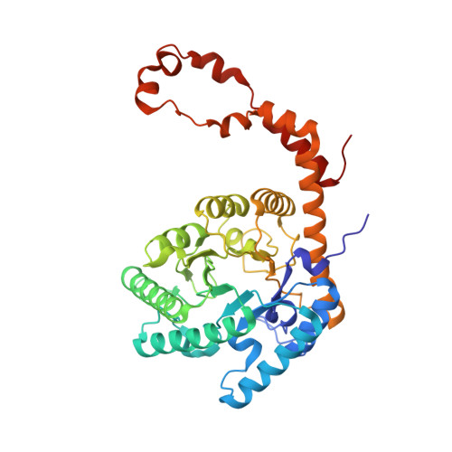
    Time-of-flight neutron diffraction has been used to locate hydrogen atoms that define the ionization states of amino acids in crystals of D-xylose isomerase. This enzyme, from Streptomyces rubiginosus, is one of the largest enzymes studied to date at high resolution (1.8 A) by this method. We have determined the position and orientation of a metal ion-bound water molecule that is located in the active site of the enzyme; this water has been thought to be involved in the isomerization step in which D-xylose is converted to D-xylulose or D-glucose to D-fructose. It is shown to be water (rather than a hydroxyl group) under the conditions of measurement (pH 8.0). Our analyses also reveal that one lysine probably has an -NH(2)-terminal group (rather than NH(3)(+)). The ionization state of each histidine residue also was determined. High-resolution x-ray studies (at 0.94 A) indicate disorder in some side chains when a truncated substrate is bound and suggest how some side chains might move during catalysis. This combination of time-of-flight neutron diffraction and x-ray diffraction can contribute greatly to the elucidation of enzyme mechanisms.


  • Organizational Affiliation

    Fox Chase Cancer Center, 333 Cottman Avenue, Philadelphia, PA 19111, USA.


Macromolecules
Find similar proteins by:  (by identity cutoff)  |  3D Structure
Entity ID: 1
MoleculeChains Sequence LengthOrganismDetailsImage
Xylose isomerase388Streptomyces rubiginosusMutation(s): 0 
EC: 5.3.1.5
UniProt
Find proteins for P24300 (Streptomyces rubiginosus)
Explore P24300 
Go to UniProtKB:  P24300
Entity Groups  
Sequence Clusters30% Identity50% Identity70% Identity90% Identity95% Identity100% Identity
UniProt GroupP24300
Sequence Annotations
Expand
  • Reference Sequence
Small Molecules
Ligands 1 Unique
IDChains Name / Formula / InChI Key2D Diagram3D Interactions
CO
Query on CO

Download Ideal Coordinates CCD File 
B [auth A],
C [auth A]
COBALT (II) ION
Co
XLJKHNWPARRRJB-UHFFFAOYSA-N
Experimental Data & Validation

Experimental Data

  • Method: NEUTRON DIFFRACTION
  • Resolution: 2.20 Å
  • R-Value Free: 0.319 
  • R-Value Work: 0.268 
  • R-Value Observed: 0.271 
  • Space Group: I 2 2 2
Unit Cell:
Length ( Å )Angle ( ˚ )
a = 93.88α = 90
b = 99.68β = 90
c = 102.9γ = 90
Software Package:
Software NamePurpose
d*TREKdata reduction
SHELXL-97model building
SHELXL-97refinement
d*TREKdata scaling
SHELXL-97phasing

Structure Validation

View Full Validation Report


Entry History 

Deposition Data

Revision History  (Full details and data files)

  • Version 1.0: 2006-05-16
    Type: Initial release
  • Version 1.1: 2008-05-01
    Changes: Version format compliance
  • Version 1.2: 2011-07-13
    Changes: Atomic model, Derived calculations, Version format compliance
  • Version 1.3: 2017-10-18
    Changes: Refinement description
  • Version 1.4: 2023-08-30
    Changes: Data collection, Database references, Derived calculations, Refinement description