


Crystallization and the 1.9 angstroms of the egg-white lysozyme from a taiwanese soft-shelled turtle (trionyx sinensis wiegmann)
Siritapetawee, J., Thammasirirak, S., Yuvaniyama, J., Robinson, R.C.To be published.
Experimental Data Snapshot
wwPDB Validation 3D Report Full Report

Entity ID: 1 | |||||
|---|---|---|---|---|---|
| Molecule | Chains | Sequence Length | Organism | Details | Image |
| Lysozyme C | 131 | Pelodiscus sinensis | Mutation(s): 0 EC: 3.2.1.17 | 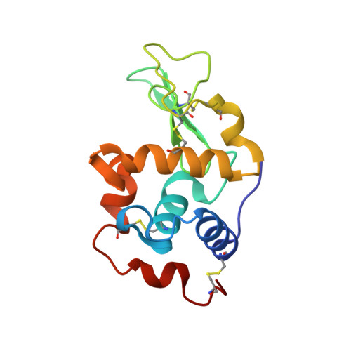 | |
UniProt | |||||
Find proteins for Q7LZQ1 (Pelodiscus sinensis) Explore Q7LZQ1 Go to UniProtKB: Q7LZQ1 | |||||
Entity Groups | |||||
| Sequence Clusters | 30% Identity50% Identity70% Identity90% Identity95% Identity100% Identity | ||||
| UniProt Group | Q7LZQ1 | ||||
Sequence AnnotationsExpand | |||||
| |||||
| Ligands 1 Unique | |||||
|---|---|---|---|---|---|
| ID | Chains | Name / Formula / InChI Key | 2D Diagram | 3D Interactions | |
| SO4 Query on SO4 | B [auth A] | SULFATE ION O4 S QAOWNCQODCNURD-UHFFFAOYSA-L |  | ||
| Length ( Å ) | Angle ( ˚ ) |
|---|---|
| a = 37.758 | α = 90 |
| b = 55.561 | β = 90 |
| c = 72.234 | γ = 90 |
| Software Name | Purpose |
|---|---|
| d*TREK | data scaling |
| AMoRE | phasing |
| REFMAC | refinement |
| PDB_EXTRACT | data extraction |
| CrystalClear | data reduction |
RCSB PDB (citation) is hosted by
RCSB PDB is a member of the

