2GU1

Crystal structure of a zinc containing peptidase from vibrio cholerae


Experimental Data Snapshot

  • Method: X-RAY DIFFRACTION
  • Resolution: 1.90 Å
  • R-Value Free: 0.264 
  • R-Value Work: 0.230 
  • R-Value Observed: 0.230 

wwPDB Validation   3D Report Full Report


This is version 1.4 of the entry. See complete history


Literature

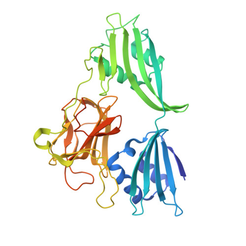
Crystal structure of a putative lysostaphin peptidase from Vibrio cholerae.

Ragumani, S.Kumaran, D.Burley, S.K.Swaminathan, S.

(2008) Proteins 72: 1096-1103


Macromolecules
Find similar proteins by:  (by identity cutoff)  |  3D Structure
Entity ID: 1
MoleculeChains Sequence LengthOrganismDetailsImage
Zinc peptidase361Vibrio choleraeMutation(s): 0 
EC: 3.4
UniProt
Find proteins for Q9KUL5 (Vibrio cholerae serotype O1 (strain ATCC 39315 / El Tor Inaba N16961))
Explore Q9KUL5 
Go to UniProtKB:  Q9KUL5
Entity Groups  
Sequence Clusters30% Identity50% Identity70% Identity90% Identity95% Identity100% Identity
UniProt GroupQ9KUL5
Sequence Annotations
Expand
  • Reference Sequence
Experimental Data & Validation

Experimental Data

  • Method: X-RAY DIFFRACTION
  • Resolution: 1.90 Å
  • R-Value Free: 0.264 
  • R-Value Work: 0.230 
  • R-Value Observed: 0.230 
  • Space Group: P 1 21 1
Unit Cell:
Length ( Å )Angle ( ˚ )
a = 49.382α = 90
b = 85.396β = 112.96
c = 53.404γ = 90
Software Package:
Software NamePurpose
CNSrefinement
HKL-2000data reduction
HKL-2000data scaling
SHELXDphasing
SHARPphasing
ARP/wARPmodel building

Structure Validation

View Full Validation Report


Entry History 

Deposition Data

Revision History  (Full details and data files)

  • Version 1.0: 2006-07-04
    Type: Initial release
  • Version 1.1: 2008-05-01
    Changes: Version format compliance
  • Version 1.2: 2011-07-13
    Changes: Version format compliance
  • Version 1.3: 2021-02-03
    Changes: Database references, Derived calculations, Structure summary
  • Version 1.4: 2024-02-14
    Changes: Data collection, Database references