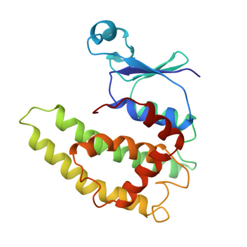
2GSQ

GLUTATHIONE S-TRANSFERASE FROM SQUID DIGESTIVE GLAND COMPLEXED WITH S-(3-IODOBENZYL)GLUTATHIONE


Experimental Data Snapshot

  • Method: X-RAY DIFFRACTION
  • Resolution: 2.20 Å
  • R-Value Observed: 0.173 

wwPDB Validation   3D Report Full Report


This is version 1.3 of the entry. See complete history


Literature

Location of a potential transport binding site in a sigma class glutathione transferase by x-ray crystallography.

Ji, X.von Rosenvinge, E.C.Johnson, W.W.Armstrong, R.N.Gilliland, G.L.

(1996) Proc Natl Acad Sci U S A 93: 8208-8213

  • DOI: https://doi.org/10.1073/pnas.93.16.8208
  • Primary Citation of Related Structures:  
    2GSQ

  • PubMed Abstract: 

    The crystal structure of the sigma class glutathione transferase from squid digestive gland in complex with S-(3-iodobenzyl)glutathione reveals a third binding site for the glutathione conjugate besides the two in the active sites of the dimer. The additional binding site is near the crystallographic two-fold axis between the two alpha 4-turn-alpha 5 motifs. The principal binding interactions with the conjugate include specific electrostatic interactions between the peptide and the two subunits and a hydrophobic cavity found across the two-fold axis that accommodates the 3-iodobenzyl group. Thus, two identical, symmetry-related but mutually exclusive binding modes for the third conjugate are observed. The hydrophobic pocket is about 14 A from the hydroxyl group of Tyr-7 in the active site. This site is a potential transport binding site for hydrophobic molecules or their glutathione conjugates.


  • Organizational Affiliation

    Center for Advanced Research in Biotechnology, University of Maryland Biotechnology Institute, Rockville, USA.


Macromolecules
Find similar proteins by:  (by identity cutoff)  |  3D Structure
Entity ID: 1
MoleculeChains Sequence LengthOrganismDetailsImage
GLUTATHIONE S-TRANSFERASE202Nototodarus sloaniiMutation(s): 0 
Gene Names: CDNA INSERT OF CLONE PGST5
EC: 2.5.1.18
UniProt
Find proteins for P46088 (Nototodarus sloanii)
Explore P46088 
Go to UniProtKB:  P46088
Entity Groups  
Sequence Clusters30% Identity50% Identity70% Identity90% Identity95% Identity100% Identity
UniProt GroupP46088
Sequence Annotations
Expand
  • Reference Sequence
Small Molecules
Ligands 2 Unique
IDChains Name / Formula / InChI Key2D Diagram3D Interactions
GBI
Query on GBI

Download Ideal Coordinates CCD File 
C [auth A],
D [auth A]
S-(3-IODOBENZYL)GLUTATHIONE
C17 H22 I N3 O6 S
AHWSFXKKIDTZBI-STQMWFEESA-N
SO4
Query on SO4

Download Ideal Coordinates CCD File 
B [auth A]SULFATE ION
O4 S
QAOWNCQODCNURD-UHFFFAOYSA-L
Experimental Data & Validation

Experimental Data

  • Method: X-RAY DIFFRACTION
  • Resolution: 2.20 Å
  • R-Value Observed: 0.173 
  • Space Group: P 31 2 1
Unit Cell:
Length ( Å )Angle ( ˚ )
a = 73.15α = 90
b = 73.15β = 90
c = 94.88γ = 120
Software Package:
Software NamePurpose
PROLSQrefinement
XENGENdata reduction

Structure Validation

View Full Validation Report


Entry History 

Deposition Data

Revision History  (Full details and data files)

  • Version 1.0: 1996-04-12
    Type: Initial release
  • Version 1.1: 2008-03-24
    Changes: Version format compliance
  • Version 1.2: 2011-07-13
    Changes: Derived calculations, Version format compliance
  • Version 1.3: 2023-08-30
    Changes: Data collection, Database references, Derived calculations, Other, Structure summary