Hijacking of a Substrate-binding Protein Scaffold for use in Mycobacterial Cell Wall Biosynthesis
Marland, Z., Beddoe, T., Zaker-Tabrizi, L., Lucet, I.S., Brammananth, R., Whisstock, J.C., Wilce, M.C., Coppel, R.L., Crellin, P.K., Rossjohn, J.(2006) J Mol Biol 359: 983-997
- PubMed: 16698034
- DOI: https://doi.org/10.1016/j.jmb.2006.04.012
- Primary Citation of Related Structures:
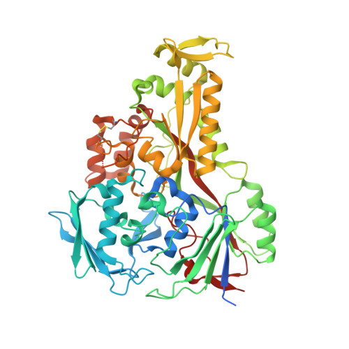
2GRV - PubMed Abstract:
The waxy cell wall is crucial to the survival of mycobacteria within the infected host. The cell wall is a complex structure rich in unusual molecules that includes two related lipoglycans, the phosphatidylinositol mannosides (PIMs) and lipoarabinomannans (LAMs). Many proteins implicated in the PIM/LAM biosynthetic pathway, while attractive therapeutic targets, are poorly defined. The 2.4A resolution crystal structure of an essential lipoprotein, LpqW, implicated in LAM biosynthesis is reported here. LpqW adopts a scaffold reminiscent of the distantly related, promiscuous substrate-binding proteins of the ATP-binding cassette import system. Nevertheless, the unique closed conformation of LpqW suggests that mycobacteria and other closely related pathogens have hijacked this scaffold for use in key processes of cell wall biosynthesis. In silico docking provided a plausible model in which the candidate PIM ligand binds within a marked electronegative region located on the surface of LpqW. We suggest that LpqW represents an archetypal lipoprotein that channels intermediates from a pathway for mature PIM production into a pathway for LAM biosynthesis, thus controlling the relative abundance of these two important components of the cell wall.
Organizational Affiliation:
The Protein Crystallography Unit, Department of Biochemistry and Molecular Biology, School of Biomedical Sciences, Monash University, Clayton, Vic. 3800, Australia.