2GQ1

Crystal Structure of Recombinant Type I Fructose-1,6-bisphosphatase from Escherichia coli Complexed with Sulfate Ions


Experimental Data Snapshot

  • Method: X-RAY DIFFRACTION
  • Resolution: 1.45 Å
  • R-Value Free: 0.232 
  • R-Value Work: 0.219 
  • R-Value Observed: 0.222 

wwPDB Validation   3D Report Full Report


This is version 1.5 of the entry. See complete history


Literature

Novel Allosteric Activation Site in Escherichia coli Fructose-1,6-bisphosphatase.

Hines, J.K.Fromm, H.J.Honzatko, R.B.

(2006) J Biol Chem 281: 18386-18393

  • DOI: https://doi.org/10.1074/jbc.M602553200
  • Primary Citation of Related Structures:  
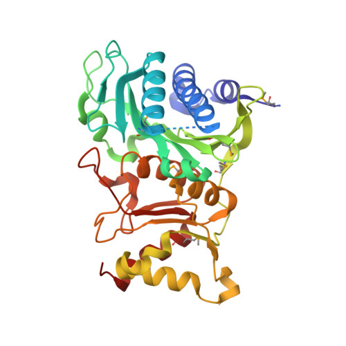
    2GQ1

  • PubMed Abstract: 

    Fructose-1,6-bisphosphatase (FBPase) governs a key step in gluconeogenesis, the conversion of fructose 1,6-bisphosphate into fructose 6-phosphate. In mammals, the enzyme is subject to metabolic regulation, but regulatory mechanisms of bacterial FBPases are not well understood. Presented here is the crystal structure (resolution, 1.45A) of recombinant FBPase from Escherichia coli, the first structure of a prokaryotic Type I FBPase. The E. coli enzyme is a homotetramer, but in a quaternary state between the canonical R- and T-states of porcine FBPase. Phe(15) and residues at the C-terminal side of the first alpha-helix (helix H1) occupy the AMP binding pocket. Residues at the N-terminal side of helix H1 hydrogen bond with sulfate ions buried at a subunit interface, which in porcine FBPase undergoes significant conformational change in response to allosteric effectors. Phosphoenolpyruvate and sulfate activate E. coli FBPase by at least 300%. Key residues that bind sulfate anions are conserved among many heterotrophic bacteria, but are absent in FBPases of organisms that employ fructose 2,6-bisphosphate as a regulator. These observations suggest a new mechanism of regulation in the FBPase enzyme family: anionic ligands, most likely phosphoenolpyruvate, bind to allosteric activator sites, which in turn stabilize a tetramer and a polypeptide fold that obstructs AMP binding.


  • Organizational Affiliation

    Department of Biochemistry, Biophysics, and Molecular Biology, Iowa State University, Ames, Iowa 50011, USA.


Macromolecules
Find similar proteins by:  (by identity cutoff)  |  3D Structure
Entity ID: 1
MoleculeChains Sequence LengthOrganismDetailsImage
Fructose-1,6-bisphosphatase332Escherichia coliMutation(s): 6 
Gene Names: fbp
EC: 3.1.3.11
UniProt
Find proteins for P0A993 (Escherichia coli (strain K12))
Explore P0A993 
Go to UniProtKB:  P0A993
Entity Groups  
Sequence Clusters30% Identity50% Identity70% Identity90% Identity95% Identity100% Identity
UniProt GroupP0A993
Sequence Annotations
Expand
  • Reference Sequence
Experimental Data & Validation

Experimental Data

  • Method: X-RAY DIFFRACTION
  • Resolution: 1.45 Å
  • R-Value Free: 0.232 
  • R-Value Work: 0.219 
  • R-Value Observed: 0.222 
  • Space Group: I 2 2 2
Unit Cell:
Length ( Å )Angle ( ˚ )
a = 45.59α = 90
b = 81.27β = 90
c = 170.1γ = 90
Software Package:
Software NamePurpose
CNSrefinement
PDB_EXTRACTdata extraction
CrystalCleardata reduction
d*TREKdata scaling
AMoREphasing
REFMACrefinement

Structure Validation

View Full Validation Report


Entry History 

Deposition Data

Revision History  (Full details and data files)

  • Version 1.0: 2006-05-02
    Type: Initial release
  • Version 1.1: 2008-05-01
    Changes: Version format compliance
  • Version 1.2: 2011-07-13
    Changes: Derived calculations, Version format compliance
  • Version 1.3: 2019-07-24
    Changes: Data collection, Derived calculations, Refinement description
  • Version 1.4: 2023-08-30
    Changes: Data collection, Database references, Derived calculations, Refinement description
  • Version 1.5: 2023-11-15
    Changes: Data collection