2GOO

Ternary Complex of BMP-2 bound to BMPR-Ia-ECD and ActRII-ECD


Experimental Data Snapshot

  • Method: X-RAY DIFFRACTION
  • Resolution: 2.20 Å
  • R-Value Free: 0.259 
  • R-Value Work: 0.222 
  • R-Value Observed: 0.224 

wwPDB Validation   3D Report Full Report


Ligand Structure Quality Assessment 


This is version 2.0 of the entry. See complete history


Literature

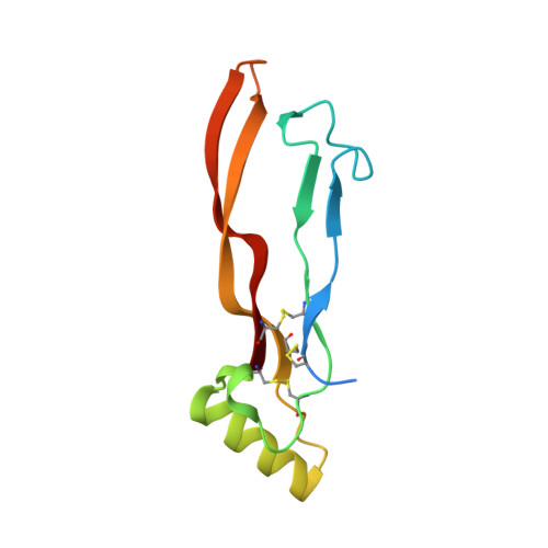
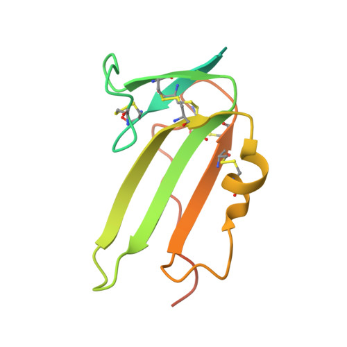
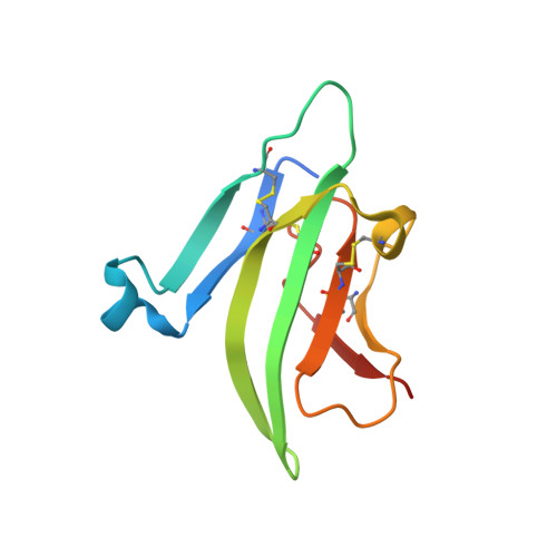
Structure of the ternary signaling complex of a TGF-beta superfamily member.

Allendorph, G.P.Vale, W.W.Choe, S.

(2006) Proc Natl Acad Sci U S A 103: 7643-7648

  • DOI: https://doi.org/10.1073/pnas.0602558103
  • Primary Citation of Related Structures:  
    2GOO

  • PubMed Abstract: 

    The crystal structure of the complete signaling complex formed between bone morphogenetic protein 2 (BMP-2) and the extracellular domains (ECDs) of its type I receptor [bone morphogenetic protein receptor type Ia (BMPR-Ia)-ECD] and its type II receptor [activin receptor type II (ActRII)-ECD] shows two fundamental structural constraints for receptor assembly. First, the homodimeric BMP-2 ligand assembles two pairs of each receptor symmetrically, where each of the receptor ECDs does not make physical contact. Therefore, conformational communication between receptor ECDs, if any, should be propagated through the central ligand. Second, the type I and II receptor interfaces of the complex, when compared with those of binary complexes such as BMP-2/BMPR Ia-ECD, BMP-7/ActRII-ECD, and activin/ActRIIb-ECD, respectively, show there are common sets of positions repeatedly used by both ligands and receptors. Therefore, specificity-determining amino acid differences at the receptor interfaces should also account for the disparity in affinity of individual receptors for different ligand subunits. We find that a specific mutation to BMP-2 increases its affinity to ActRII-ECD by 5-fold. These results together establish that the specific signaling output is largely determined by two variables, the ligand-receptor pair identity and the mode of cooperative assembly of relevant receptors governed by the ligand flexibility in a membrane-restricted manner.


  • Organizational Affiliation

    Structural Biology Laboratory, The Salk Institute, La Jolla, CA 92037, USA.


Macromolecules
Find similar proteins by:  (by identity cutoff)  |  3D Structure
Entity ID: 1
MoleculeChains Sequence LengthOrganismDetailsImage
Bone morphogenetic protein 2
A, D
114Homo sapiensMutation(s): 0 
Gene Names: BMP2
UniProt & NIH Common Fund Data Resources
Find proteins for P12643 (Homo sapiens)
Explore P12643 
Go to UniProtKB:  P12643
PHAROS:  P12643
GTEx:  ENSG00000125845 
Entity Groups  
Sequence Clusters30% Identity50% Identity70% Identity90% Identity95% Identity100% Identity
UniProt GroupP12643
Sequence Annotations
Expand
  • Reference Sequence
Find similar proteins by:  (by identity cutoff)  |  3D Structure
Entity ID: 2
MoleculeChains Sequence LengthOrganismDetailsImage
Bone morphogenetic protein receptor type IA
B, E
131Homo sapiensMutation(s): 0 
Gene Names: BMPR1A
EC: 2.7.11.30
UniProt & NIH Common Fund Data Resources
Find proteins for P36894 (Homo sapiens)
Explore P36894 
Go to UniProtKB:  P36894
PHAROS:  P36894
GTEx:  ENSG00000107779 
Entity Groups  
Sequence Clusters30% Identity50% Identity70% Identity90% Identity95% Identity100% Identity
UniProt GroupP36894
Sequence Annotations
Expand
  • Reference Sequence
Find similar proteins by:  (by identity cutoff)  |  3D Structure
Entity ID: 3
MoleculeChains Sequence LengthOrganismDetailsImage
Activin receptor type 2A
C, F
102Mus musculusMutation(s): 0 
Gene Names: Acvr2a
EC: 2.7.11.30
UniProt & NIH Common Fund Data Resources
Find proteins for P27038 (Mus musculus)
Explore P27038 
Go to UniProtKB:  P27038
IMPC:  MGI:102806
Entity Groups  
Sequence Clusters30% Identity50% Identity70% Identity90% Identity95% Identity100% Identity
UniProt GroupP27038
Sequence Annotations
Expand
  • Reference Sequence
Experimental Data & Validation

Experimental Data

  • Method: X-RAY DIFFRACTION
  • Resolution: 2.20 Å
  • R-Value Free: 0.259 
  • R-Value Work: 0.222 
  • R-Value Observed: 0.224 
  • Space Group: P 65 2 2
Unit Cell:
Length ( Å )Angle ( ˚ )
a = 104.031α = 90
b = 104.031β = 90
c = 362.526γ = 120
Software Package:
Software NamePurpose
DENZOdata reduction
SCALEPACKdata scaling
REFMACrefinement
PDB_EXTRACTdata extraction
Blu-Icedata collection
HKL-2000data scaling
PHASERphasing

Structure Validation

View Full Validation Report


Ligand Structure Quality Assessment 


Entry History 

Deposition Data

Revision History  (Full details and data files)

  • Version 1.0: 2006-05-09
    Type: Initial release
  • Version 1.1: 2008-05-01
    Changes: Version format compliance
  • Version 1.2: 2011-07-13
    Changes: Version format compliance
  • Version 1.3: 2017-10-18
    Changes: Advisory, Refinement description
  • Version 2.0: 2020-07-29
    Type: Remediation
    Reason: Carbohydrate remediation
    Changes: Advisory, Atomic model, Data collection, Database references, Derived calculations, Structure summary