Structural Basis of Robo Proline-rich Motif Recognition by the srGAP1 Src Homology 3 Domain in the Slit-Robo Signaling Pathway
Li, X., Chen, Y., Liu, Y., Gao, J., Gao, F., Bartlam, M., Wu, J.Y., Rao, Z.(2006) J Biol Chem 281: 28430-28437
- PubMed: 16857672
- DOI: https://doi.org/10.1074/jbc.M604135200
- Primary Citation of Related Structures:
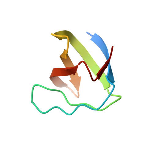
2GNC - PubMed Abstract:
The Slit-Robo (sr) GTPase-activating protein (GAPs) are important components in the intracellular pathway mediating Slit-Robo signaling in axon guidance and cell migration. We report the first crystal structure of the srGAP1 SH3 domain at 1.8-A resolution. The unusual side chain conformation of the conserved Phe-13 in the P1 pocket renders the ligand binding pocket shallow and narrow, which contributes toward the low binding affinity. Moreover, the opposing electrostatic charge and the hydrophobic properties of the P3 specificity pocket are consistent with the observed binding characteristics of the srGAP1 SH3 domain to its ligand. Surface plasmon resonance experiments indicate that the srGAP1 SH3 domain interacts with its natural ligand inaCtoN orientation. The srGAP1 SH3 domain can bind to both the CC2 and CC3 motifs in vitro. The N-terminal two acidic residues in the CC3 motif recognition site are necessary for srGAP1 SH3 domain binding. A longer CC3 peptide (CC3-FL) binds with greater affinity than its shorter counterpart, suggesting that the residues surrounding the proline-rich core are important for protein-peptide interactions. Our study reveals previously unknown properties of the srGAP-Robo interaction. Our data provide a structural basis for the srGAP-Robo interaction, consistent with the role of the Robo intracellular domain in interacting with other downstream signaling molecules and mediating versatile and dynamic responses to axon guidance and cell migration cues.
Organizational Affiliation:
Tsinghua-Nankai IBP Joint Research Group for Structural Biology, Tsinghua University, Beijing 100084, China.