2GH5

Crystal Structure of human Glutathione Reductase complexed with a Fluoro-Analogue of the Menadione Derivative M5


Experimental Data Snapshot

  • Method: X-RAY DIFFRACTION
  • Resolution: 1.70 Å
  • R-Value Free: 0.256 
  • R-Value Work: 0.224 
  • R-Value Observed: 0.224 

wwPDB Validation   3D Report Full Report


Ligand Structure Quality Assessment 


This is version 1.3 of the entry. See complete history


Literature

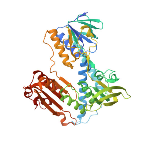
A fluoro analogue of the menadione derivative 6-[2'-(3'-methyl)-1',4'-naphthoquinolyl]hexanoic acid is a suicide substrate of glutathione reductase. Crystal structure of the alkylated human enzyme

Bauer, H.Fritz-Wolf, K.Winzer, A.Little, S.Yardley, V.Vezin, H.Palfey, B.Schirmer, R.H.Davioud-Charvet, E.

(2006) J Am Chem Soc 128: 10784-10794

  • DOI: https://doi.org/10.1021/ja061155v
  • Primary Citation of Related Structures:  
    2GH5

  • PubMed Abstract: 

    Glutathione reductase is an important housekeeping enzyme for redox homeostasis both in human cells and in the causative agent of tropical malaria, Plasmodium falciparum. Glutathione reductase inhibitors were shown to have anticancer and antimalarial activity per se and to contribute to the reversal of drug resistance. The development of menadione chemistry has led to the selection of 6-[2'-(3'-methyl)-1',4'-naphthoquinolyl]hexanoic acid, called M(5), as a potent reversible and uncompetitive inhibitor of both human and P. falciparum glutathione reductases. Here we describe the synthesis and kinetic characterization of a fluoromethyl-M(5) analogue that acts as a mechanism-based inhibitor of both enzymes. In the course of enzymatic catalysis, the suicide substrate is activated by one- or two-electron reduction, and then a highly reactive quinone methide is generated upon elimination of the fluorine. Accordingly the human enzyme was found to be irreversibly inactivated with a k(inact) value of 0.4 +/- 0.2 min(-1). The crystal structure of the alkylated enzyme was solved at 1.7 A resolution. It showed the inhibitor to bind covalently to the active site Cys58 and to interact noncovalently with His467', Arg347, Arg37, and Tyr114. On the basis of the crystal structure of the inactivated human enzyme and stopped-flow kinetic studies with two- and four-electron-reduced forms of the unreacted P. falciparum enzyme, a mechanism is proposed which explains naphthoquinone reduction at the flavin of glutathione reductase.


  • Organizational Affiliation

    Biochemie-Zentrum der Universität Heidelberg, Im Neuenheimer Feld 504, D-69120 Heidelberg, Germany.


Macromolecules
Find similar proteins by:  (by identity cutoff)  |  3D Structure
Entity ID: 1
MoleculeChains Sequence LengthOrganismDetailsImage
glutathione reductase, mitochondrial
A, B
478Homo sapiensMutation(s): 0 
Gene Names: GSRGLURGRD1
EC: 1.8.1.7
UniProt & NIH Common Fund Data Resources
Find proteins for P00390 (Homo sapiens)
Explore P00390 
Go to UniProtKB:  P00390
PHAROS:  P00390
GTEx:  ENSG00000104687 
Entity Groups  
Sequence Clusters30% Identity50% Identity70% Identity90% Identity95% Identity100% Identity
UniProt GroupP00390
Sequence Annotations
Expand
  • Reference Sequence
Small Molecules
Ligands 4 Unique
IDChains Name / Formula / InChI Key2D Diagram3D Interactions
FAD
Query on FAD

Download Ideal Coordinates CCD File 
F [auth A],
O [auth B]
FLAVIN-ADENINE DINUCLEOTIDE
C27 H33 N9 O15 P2
VWWQXMAJTJZDQX-UYBVJOGSSA-N
ELI
Query on ELI

Download Ideal Coordinates CCD File 
E [auth A],
N [auth B]
6-(3-METHYL-1,4-DIOXO-1,4-DIHYDRONAPHTHALEN-2-YL)HEXANOIC ACID
C17 H18 O4
ICGRXHWXPCXIKM-UHFFFAOYSA-N
PO4
Query on PO4

Download Ideal Coordinates CCD File 
C [auth A],
D [auth A],
L [auth B],
M [auth B]
PHOSPHATE ION
O4 P
NBIIXXVUZAFLBC-UHFFFAOYSA-K
GOL
Query on GOL

Download Ideal Coordinates CCD File 
G [auth A]
H [auth A]
I [auth A]
J [auth A]
K [auth A]
G [auth A],
H [auth A],
I [auth A],
J [auth A],
K [auth A],
P [auth B],
Q [auth B],
R [auth B],
S [auth B],
T [auth B],
U [auth B],
V [auth B]
GLYCEROL
C3 H8 O3
PEDCQBHIVMGVHV-UHFFFAOYSA-N
Experimental Data & Validation

Experimental Data

  • Method: X-RAY DIFFRACTION
  • Resolution: 1.70 Å
  • R-Value Free: 0.256 
  • R-Value Work: 0.224 
  • R-Value Observed: 0.224 
  • Space Group: P 1 21 1
Unit Cell:
Length ( Å )Angle ( ˚ )
a = 84.7α = 90
b = 63.6β = 101.12
c = 103.99γ = 90
Software Package:
Software NamePurpose
XDSdata reduction
CNSrefinement
XDSdata scaling
CNSphasing

Structure Validation

View Full Validation Report


Ligand Structure Quality Assessment 


Entry History 

Deposition Data

Revision History  (Full details and data files)

  • Version 1.0: 2006-09-26
    Type: Initial release
  • Version 1.1: 2008-05-01
    Changes: Version format compliance
  • Version 1.2: 2011-07-13
    Changes: Non-polymer description, Version format compliance
  • Version 1.3: 2023-08-30
    Changes: Data collection, Database references, Derived calculations, Refinement description