2GFI

Crystal structure of the phytase from D. castellii at 2.3 A


Experimental Data Snapshot

  • Method: X-RAY DIFFRACTION
  • Resolution: 2.29 Å
  • R-Value Free: 0.210 
  • R-Value Work: 0.154 

wwPDB Validation   3D Report Full Report


Ligand Structure Quality Assessment 


This is version 1.4 of the entry. See complete history


Literature

Structure of Debaryomyces castellii CBS 2923 phytase.

Ragon, M.Hoh, F.Aumelas, A.Chiche, L.Moulin, G.Boze, H.

(2009) Acta Crystallogr Sect F Struct Biol Cryst Commun 65: 321-326

  • DOI: https://doi.org/10.1107/S1744309109008653
  • Primary Citation of Related Structures:  
    2GFI

  • PubMed Abstract: 

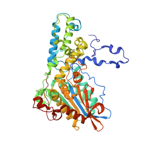
    Phytate (myo-inositol hexakisphosphate) is the primary storage form of phosphate in seeds and legumes (Reddy et al., 1982). Phytases are phosphatases that hydrolyze phytate to less phosphorylated myo-inositol derivatives and inorganic phosphate. The crystal structure of phytase from Debaryomyces castellii has been determined at 2.3 A resolution. The crystals belonged to space group P6(5)22, with unit-cell parameters a = 121.65, c = 332.24 A. The structure was solved by molecular replacement and refined to a final R factor of 15.7% (R(free) = 20.9%). The final model consists of a dimer (with two monomers of 458 residues), five NAG molecules and 628 water molecules.


  • Organizational Affiliation

    UMR IR2B, Equipe Génie Microbiologique et Enzymatique, ENSAM-INRA, Montpellier, France.


Macromolecules
Find similar proteins by:  (by identity cutoff)  |  3D Structure
Entity ID: 1
MoleculeChains Sequence LengthOrganismDetailsImage
phytase
A, B
458Debaryomyces castelliiMutation(s): 0 
EC: 3.1.3.8
UniProt
Find proteins for A2TBB4 (Debaryomyces castellii)
Explore A2TBB4 
Go to UniProtKB:  A2TBB4
Entity Groups  
Sequence Clusters30% Identity50% Identity70% Identity90% Identity95% Identity100% Identity
UniProt GroupA2TBB4
Sequence Annotations
Expand
  • Reference Sequence
Experimental Data & Validation

Experimental Data

  • Method: X-RAY DIFFRACTION
  • Resolution: 2.29 Å
  • R-Value Free: 0.210 
  • R-Value Work: 0.154 
  • Space Group: P 65 2 2
Unit Cell:
Length ( Å )Angle ( ˚ )
a = 121.655α = 90
b = 121.655β = 90
c = 332.245γ = 120
Software Package:
Software NamePurpose
SCALAdata scaling
REFMACrefinement
PDB_EXTRACTdata extraction
MOSFLMdata reduction
CCP4data scaling
PHASERphasing

Structure Validation

View Full Validation Report


Ligand Structure Quality Assessment 


Entry History 

Deposition Data

  • Released Date: 2007-03-27 
  • Deposition Author(s): Hoh, F.

Revision History  (Full details and data files)

  • Version 1.0: 2007-03-27
    Type: Initial release
  • Version 1.1: 2008-04-01
    Changes: Version format compliance
  • Version 1.2: 2011-07-13
    Changes: Derived calculations, Version format compliance
  • Version 1.3: 2020-07-29
    Type: Remediation
    Reason: Carbohydrate remediation
    Changes: Data collection, Derived calculations, Structure summary
  • Version 1.4: 2023-08-30
    Changes: Data collection, Database references, Refinement description, Structure summary