2GFA

double tudor domain complex structure


Experimental Data Snapshot

  • Method: X-RAY DIFFRACTION
  • Resolution: 2.10 Å
  • R-Value Free: 0.270 
  • R-Value Work: 0.224 
  • R-Value Observed: 0.270 

wwPDB Validation   3D Report Full Report


This is version 1.4 of the entry. See complete history


Literature

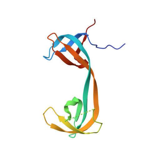
Recognition of histone H3 lysine-4 methylation by the double tudor domain of JMJD2A

Huang, Y.Fang, J.Bedford, M.T.Zhang, Y.Xu, R.M.

(2006) Science 312: 748-751

  • DOI: https://doi.org/10.1126/science.1125162
  • Primary Citation of Related Structures:  
    2GF7, 2GFA

  • PubMed Abstract: 

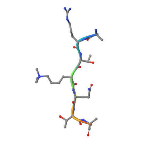
    Biological responses to histone methylation critically depend on the faithful readout and transduction of the methyl-lysine signal by "effector" proteins, yet our understanding of methyl-lysine recognition has so far been limited to the study of histone binding by chromodomain and WD40-repeat proteins. The double tudor domain of JMJD2A, a Jmjc domain-containing histone demethylase, binds methylated histone H3-K4 and H4-K20. We found that the double tudor domain has an interdigitated structure, and the unusual fold is required for its ability to bind methylated histone tails. The cocrystal structure of the JMJD2A double tudor domain with a trimethylated H3-K4 peptide reveals that the trimethyl-K4 is bound in a cage of three aromatic residues, two of which are from the tudor-2 motif, whereas the binding specificity is determined by side-chain interactions involving amino acids from the tudor-1 motif. Our study provides mechanistic insights into recognition of methylated histone tails by tudor domains and reveals the structural intricacy of methyl-lysine recognition by two closely spaced effector domains.


  • Organizational Affiliation

    W. M. Keck Structural Biology Laboratory, Cold Spring Harbor Laboratory, Cold Spring Harbor, NY 11724, USA.


Macromolecules
Find similar proteins by:  (by identity cutoff)  |  3D Structure
Entity ID: 1
MoleculeChains Sequence LengthOrganismDetailsImage
Jumonji domain-containing protein 2A
A, B
119Homo sapiensMutation(s): 0 
Gene Names: JMJD2AJMJD2KIAA0677
UniProt & NIH Common Fund Data Resources
Find proteins for O75164 (Homo sapiens)
Explore O75164 
Go to UniProtKB:  O75164
PHAROS:  O75164
GTEx:  ENSG00000066135 
Entity Groups  
Sequence Clusters30% Identity50% Identity70% Identity90% Identity95% Identity100% Identity
UniProt GroupO75164
Sequence Annotations
Expand
  • Reference Sequence

Find similar proteins by:  Sequence   |   3D Structure  

Entity ID: 2
MoleculeChains Sequence LengthOrganismDetailsImage
peptide
C, D
10N/AMutation(s): 0 
Entity Groups  
Sequence Clusters30% Identity50% Identity70% Identity90% Identity95% Identity100% Identity
Sequence Annotations
Expand
  • Reference Sequence
Small Molecules
Modified Residues  1 Unique
IDChains TypeFormula2D DiagramParent
M3L
Query on M3L
C, D
L-PEPTIDE LINKINGC9 H21 N2 O2LYS
Experimental Data & Validation

Experimental Data

  • Method: X-RAY DIFFRACTION
  • Resolution: 2.10 Å
  • R-Value Free: 0.270 
  • R-Value Work: 0.224 
  • R-Value Observed: 0.270 
  • Space Group: C 2 2 21
Unit Cell:
Length ( Å )Angle ( ˚ )
a = 63.822α = 90
b = 96.733β = 90
c = 110.057γ = 90
Software Package:
Software NamePurpose
DENZOdata reduction
SCALEPACKdata scaling
PHASERphasing
CNSrefinement
PDB_EXTRACTdata extraction
ADSCdata collection

Structure Validation

View Full Validation Report


Entry History 

Deposition Data

Revision History  (Full details and data files)

  • Version 1.0: 2006-05-02
    Type: Initial release
  • Version 1.1: 2008-05-01
    Changes: Version format compliance
  • Version 1.2: 2011-07-13
    Changes: Version format compliance
  • Version 1.3: 2017-10-18
    Changes: Refinement description
  • Version 1.4: 2023-08-30
    Changes: Data collection, Database references, Derived calculations, Refinement description