2GDZ

Crystal structure of 15-hydroxyprostaglandin dehydrogenase type1, complexed with NAD+


Experimental Data Snapshot

  • Method: X-RAY DIFFRACTION
  • Resolution: 1.65 Å
  • R-Value Free: 0.212 
  • R-Value Work: 0.176 
  • R-Value Observed: 0.177 

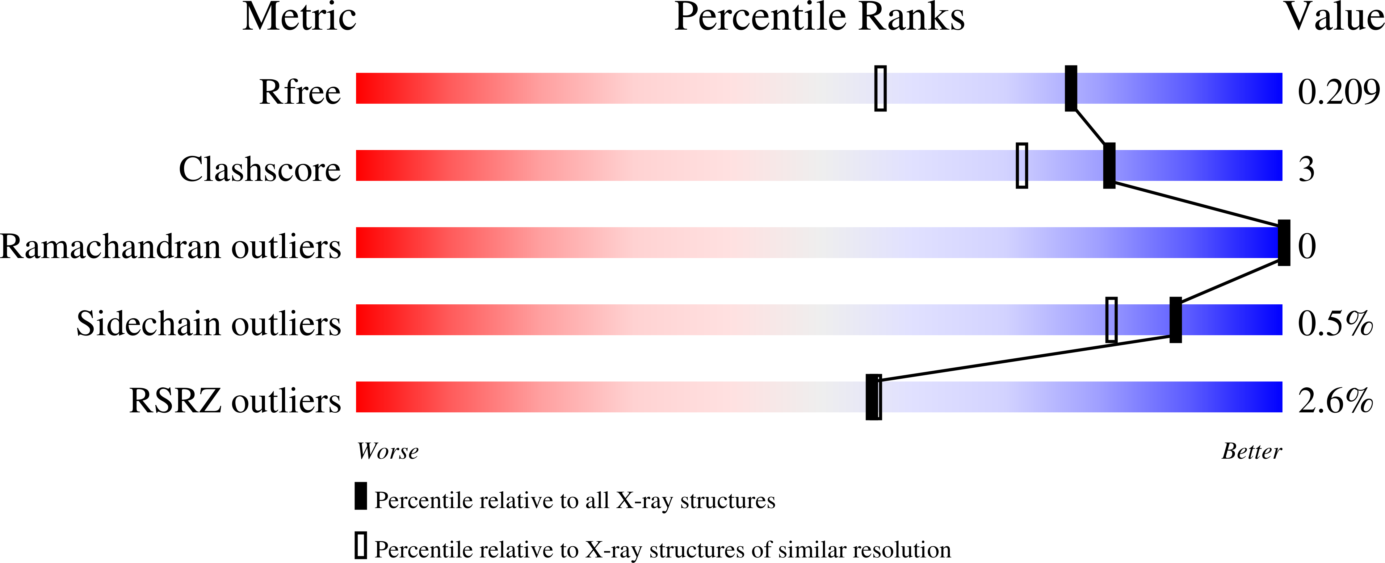
wwPDB Validation   3D Report Full Report


Ligand Structure Quality Assessment 


This is version 1.3 of the entry. See complete history


Literature

High-Affinity Inhibitors of Human NAD-Dependent 15-Hydroxyprostaglandin Dehydrogenase: Mechanisms of Inhibition and Structure-Activity Relationships.

Niesen, F.H.Schultz, L.Jadhav, A.Bhatia, C.Guo, K.Maloney, D.J.Pilka, E.S.Wang, M.Oppermann, U.Heightman, T.D.Simeonov, A.

(2010) PLoS One 5: e13719-e13719

  • DOI: https://doi.org/10.1371/journal.pone.0013719
  • Primary Citation of Related Structures:  
    2GDZ

  • PubMed Abstract: 

    15-Hydroxyprostaglandin dehydrogenase (15-PGDH, EC 1.1.1.141) is the key enzyme for the inactivation of prostaglandins, regulating processes such as inflammation or proliferation. The anabolic pathways of prostaglandins, especially with respect to regulation of the cyclooxygenase (COX) enzymes have been studied in detail; however, little is known about downstream events including functional interaction of prostaglandin-processing and -metabolizing enzymes. High-affinity probes for 15-PGDH will, therefore, represent important tools for further studies.


  • Organizational Affiliation

    Structural Genomics Consortium, Nuffield Department of Clinical Medicine, University of Oxford, Oxford, United Kingdom.


Macromolecules
Find similar proteins by:  (by identity cutoff)  |  3D Structure
Entity ID: 1
MoleculeChains Sequence LengthOrganismDetailsImage
NAD+-dependent 15-hydroxyprostaglandin dehydrogenase267Homo sapiensMutation(s): 0 
EC: 1.1.1.141
UniProt & NIH Common Fund Data Resources
Find proteins for P15428 (Homo sapiens)
Explore P15428 
Go to UniProtKB:  P15428
PHAROS:  P15428
GTEx:  ENSG00000164120 
Entity Groups  
Sequence Clusters30% Identity50% Identity70% Identity90% Identity95% Identity100% Identity
UniProt GroupP15428
Sequence Annotations
Expand
  • Reference Sequence
Small Molecules
Ligands 1 Unique
IDChains Name / Formula / InChI Key2D Diagram3D Interactions
NAD
Query on NAD

Download Ideal Coordinates CCD File 
B [auth A]NICOTINAMIDE-ADENINE-DINUCLEOTIDE
C21 H27 N7 O14 P2
BAWFJGJZGIEFAR-NNYOXOHSSA-N
Experimental Data & Validation

Experimental Data

  • Method: X-RAY DIFFRACTION
  • Resolution: 1.65 Å
  • R-Value Free: 0.212 
  • R-Value Work: 0.176 
  • R-Value Observed: 0.177 
  • Space Group: P 41 21 2
Unit Cell:
Length ( Å )Angle ( ˚ )
a = 49.54α = 90
b = 49.54β = 90
c = 195.771γ = 90
Software Package:
Software NamePurpose
REFMACrefinement
MOSFLMdata reduction
CCP4data scaling
PHASERphasing

Structure Validation

View Full Validation Report


Ligand Structure Quality Assessment 


Entry History 

Revision History  (Full details and data files)

  • Version 1.0: 2006-04-04
    Type: Initial release
  • Version 1.1: 2008-05-01
    Changes: Version format compliance
  • Version 1.2: 2011-07-13
    Changes: Derived calculations, Version format compliance
  • Version 1.3: 2023-10-25
    Changes: Data collection, Database references, Derived calculations, Refinement description