2GDS

Interrupting the Hydrogen Bonding Network at the Active Site of Human Manganese Superoxide Dismutase


Experimental Data Snapshot

  • Method: X-RAY DIFFRACTION
  • Resolution: 2.30 Å
  • R-Value Free: 0.300 
  • R-Value Work: 0.199 
  • R-Value Observed: 0.199 

wwPDB Validation   3D Report Full Report


This is version 1.4 of the entry. See complete history


Literature

Interrupting the Hydrogen Bond Network at the Active Site of Human Manganese Superoxide Dismutase

Ramilo, C.A.Leveque, V.Guan, Y.Lepock, J.R.Tainer, J.A.Nick, H.S.Silverman, D.N.

(1999) J Biol Chem 274: 27711-27716

  • DOI: https://doi.org/10.1074/jbc.274.39.27711
  • Primary Citation of Related Structures:  
    2GDS

  • PubMed Abstract: 

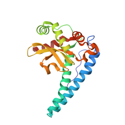
    Histidine 30 in human manganese superoxide dismutase (MnSOD) is located at a site partially exposed to solvent with its side chain participating in a hydrogen-bonded network that includes the active-site residues Tyr(166) and Tyr(34) and extends to the manganese-bound solvent molecule. We have replaced His(30) with a series of amino acids and Tyr(166) with Phe in human MnSOD. The crystal structure of the mutant of MnSOD containing Asn(30) superimposed closely with the wild type, but the side chain of Asn(30) did not participate in the hydrogen-bonded network in the active site. The catalytic activity of a number of mutants with replacements at position 30 and for the mutant containing Phe(166) showed a 10-40-fold decrease in k(cat). This is the same magnitude of decrease in k(cat) obtained with the replacement of Tyr(34) by Phe, suggesting that interrupting the hydrogen-bonded active-site network at any of the sites of these three participants (His(30), Tyr(34), and Tyr(166)) leads to an equivalent decrease in k(cat) and probably less efficient proton transfer to product peroxide. The specific geometry of His(30) on the hydrogen bond network is essential for stability since the disparate mutations H30S, H30A, and H30Q reduce T(m) by similar amounts (10-16 degrees C) compared with wild type.


  • Organizational Affiliation

    Department of Pharmacology and Therapeutics, University of Florida, Gainesville, Florida 32610, USA.


Macromolecules
Find similar proteins by:  (by identity cutoff)  |  3D Structure
Entity ID: 1
MoleculeChains Sequence LengthOrganismDetailsImage
Superoxide dismutase
A, B, C, D
198Homo sapiensMutation(s): 1 
Gene Names: SOD2
EC: 1.15.1.1
UniProt & NIH Common Fund Data Resources
Find proteins for P04179 (Homo sapiens)
Explore P04179 
Go to UniProtKB:  P04179
PHAROS:  P04179
Entity Groups  
Sequence Clusters30% Identity50% Identity70% Identity90% Identity95% Identity100% Identity
UniProt GroupP04179
Sequence Annotations
Expand
  • Reference Sequence
Experimental Data & Validation

Experimental Data

  • Method: X-RAY DIFFRACTION
  • Resolution: 2.30 Å
  • R-Value Free: 0.300 
  • R-Value Work: 0.199 
  • R-Value Observed: 0.199 
  • Space Group: P 21 21 21
Unit Cell:
Length ( Å )Angle ( ˚ )
a = 73.7α = 90
b = 77.57β = 90
c = 135.46γ = 90
Software Package:
Software NamePurpose
ADSCdata collection
DENZOdata reduction
AMoREphasing
X-PLORrefinement
SCALEPACKdata scaling

Structure Validation

View Full Validation Report


Entry History 

Deposition Data

  • Released Date: 2006-03-28 
  • Deposition Author(s): Perry, J.J.

Revision History  (Full details and data files)

  • Version 1.0: 2006-03-28
    Type: Initial release
  • Version 1.1: 2008-05-01
    Changes: Version format compliance
  • Version 1.2: 2011-07-13
    Changes: Version format compliance
  • Version 1.3: 2021-10-20
    Changes: Database references, Derived calculations
  • Version 1.4: 2023-08-30
    Changes: Data collection, Refinement description