2GDR

Crystal structure of a bacterial glutathione transferase


Experimental Data Snapshot

  • Method: X-RAY DIFFRACTION
  • Resolution: 2.10 Å
  • R-Value Free: 0.204 
  • R-Value Work: 0.161 
  • R-Value Observed: 0.161 

wwPDB Validation   3D Report Full Report


Ligand Structure Quality Assessment 


This is version 1.4 of the entry. See complete history


Literature

Structures of Ternary Complexes of BphK, a Bacterial Glutathione S-Transferase That Reductively Dechlorinates Polychlorinated Biphenyl Metabolites.

Tocheva, E.I.Fortin, P.D.Eltis, L.D.Murphy, M.E.

(2006) J Biol Chem 281: 30933-30940

  • DOI: https://doi.org/10.1074/jbc.M603125200

  • PubMed Abstract: 

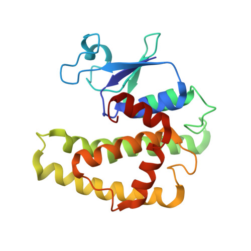
    Prokaryotic glutathione S-transferases are as diverse as their eukaryotic counterparts but are much less well characterized. BphK from Burkholderia xenovorans LB400 consumes two GSH molecules to reductively dehalogenate chlorinated 2-hydroxy-6-oxo-6-phenyl-2,4-dienoates (HOPDAs), inhibitory polychlorinated biphenyl metabolites. Crystallographic structures of two ternary complexes of BphK were solved to a resolution of 2.1A. In the BphK-GSH-HOPDA complex, GSH and HOPDA molecules occupy the G- and H-subsites, respectively. The thiol nucleophile of the GSH molecule is positioned for SN2 attack at carbon 3 of the bound HOPDA. The respective sulfur atoms of conserved Cys-10 and the bound GSH are within 3.0A, consistent with product release and the formation of a mixed disulfide intermediate. In the BphK-(GSH)2 complex, a GSH molecule occupies each of the two subsites. The three sulfur atoms of the two GSH molecules and Cys-10 are aligned suitably for a disulfide exchange reaction that would regenerate the resting enzyme and yield disulfide-linked GSH molecules. A second conserved residue, His-106, is adjacent to the thiols of Cys-10 and the GSH bound to the G-subsite and thus may stabilize a transition state in the disulfide exchange reaction. Overall, the structures support and elaborate a proposed dehalogenation mechanism for BphK and provide insight into the plasticity of the H-subsite.


  • Organizational Affiliation

    Department of Microbiology and Immunology, University of British Columbia, Vancouver, British Columbia V6T 1Z3, Canada.


Macromolecules
Find similar proteins by:  (by identity cutoff)  |  3D Structure
Entity ID: 1
MoleculeChains Sequence LengthOrganismDetailsImage
glutathione S-transferase
A, B, C, D, E
A, B, C, D, E, F
202Paraburkholderia xenovorans LB400Mutation(s): 0 
Gene Names: bphK
EC: 2.5.1.18
UniProt
Find proteins for Q59721 (Paraburkholderia xenovorans (strain LB400))
Explore Q59721 
Go to UniProtKB:  Q59721
Entity Groups  
Sequence Clusters30% Identity50% Identity70% Identity90% Identity95% Identity100% Identity
UniProt GroupQ59721
Sequence Annotations
Expand
  • Reference Sequence
Experimental Data & Validation

Experimental Data

  • Method: X-RAY DIFFRACTION
  • Resolution: 2.10 Å
  • R-Value Free: 0.204 
  • R-Value Work: 0.161 
  • R-Value Observed: 0.161 
  • Space Group: P 65
Unit Cell:
Length ( Å )Angle ( ˚ )
a = 103.44α = 90
b = 103.44β = 90
c = 262.28γ = 120
Software Package:
Software NamePurpose
DENZOdata reduction
SCALEPACKdata scaling
CNSrefinement
CNSphasing

Structure Validation

View Full Validation Report


Ligand Structure Quality Assessment 


Entry History 

Deposition Data

Revision History  (Full details and data files)

  • Version 1.0: 2006-08-22
    Type: Initial release
  • Version 1.1: 2008-05-01
    Changes: Version format compliance
  • Version 1.2: 2011-07-13
    Changes: Source and taxonomy, Version format compliance
  • Version 1.3: 2011-12-14
    Changes: Non-polymer description
  • Version 1.4: 2023-08-30
    Changes: Data collection, Database references, Derived calculations, Refinement description