2GB7

Metal-depleted Ecl18kI in complex with uncleaved, modified DNA


Experimental Data Snapshot

  • Method: X-RAY DIFFRACTION
  • Resolution: 1.70 Å
  • R-Value Free: 0.236 
  • R-Value Work: 0.207 
  • R-Value Observed: 0.208 

wwPDB Validation   3D Report Full Report


This is version 1.4 of the entry. See complete history


Literature

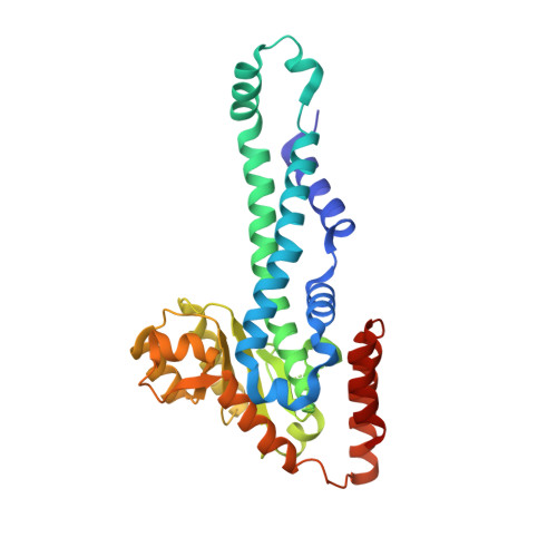
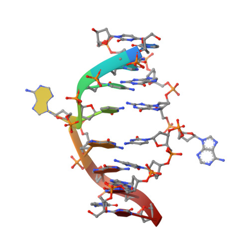
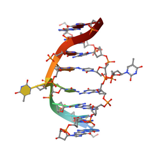
Nucleotide flips determine the specificity of the Ecl18kI restriction endonuclease

Bochtler, M.Szczepanowski, R.H.Tamulaitis, G.Grazulis, S.Czapinska, H.Manakova, E.Siksnys, V.

(2006) EMBO J 25: 2219-2229

  • DOI: https://doi.org/10.1038/sj.emboj.7601096
  • Primary Citation of Related Structures:  
    2FQZ, 2GB7

  • PubMed Abstract: 

    Restricion endonuclease Ecl18kI is specific for the sequence /CCNGG and cleaves it before the outer C to generate 5 nt 5'-overhangs. It has been suggested that Ecl18kI is evolutionarily related to NgoMIV, a 6-bp cutter that cleaves the sequence G/CCGGC and leaves 4 nt 5'-overhangs. Here, we report the crystal structure of the Ecl18kI-DNA complex at 1.7 A resolution and compare it with the known structure of the NgoMIV-DNA complex. We find that Ecl18kI flips both central nucleotides within the CCNGG sequence and buries the extruded bases in pockets within the protein. Nucleotide flipping disrupts Watson-Crick base pairing, induces a kink in the DNA and shifts the DNA register by 1 bp, making the distances between scissile phosphates in the Ecl18kI and NgoMIV cocrystal structures nearly identical. Therefore, the two enzymes can use a conserved DNA recognition module, yet recognize different sequences, and form superimposable dimers, yet generate different cleavage patterns. Hence, Ecl18kI is the first example of a restriction endonuclease that flips nucleotides to achieve specificity for its recognition site.


  • Organizational Affiliation

    International Institute of Molecular and Cell Biology, Warsaw, Poland. MBochtler@iimcb.gov.pl


Macromolecules

Find similar proteins by:  (by identity cutoff)  |  3D Structure
Entity ID: 3
MoleculeChains Sequence LengthOrganismDetailsImage
R.Ecl18kIE [auth A],
F [auth B],
G [auth C],
H [auth D]
305Enterobacter cloacaeMutation(s): 1 
Gene Names: ecl18kIR
UniProt
Find proteins for O87963 (Enterobacter cloacae)
Explore O87963 
Go to UniProtKB:  O87963
Entity Groups  
Sequence Clusters30% Identity50% Identity70% Identity90% Identity95% Identity100% Identity
UniProt GroupO87963
Sequence Annotations
Expand
  • Reference Sequence

Find similar nucleic acids by:  Sequence   |   3D Structure  

Entity ID: 1
MoleculeChains LengthOrganismImage
DNA STRAND 1A [auth E],
C [auth G]
9N/A
Sequence Annotations
Expand
  • Reference Sequence

Find similar nucleic acids by:  Sequence   |   3D Structure  

Entity ID: 2
MoleculeChains LengthOrganismImage
DNA STRAND 2B [auth F],
D [auth H]
9N/A
Sequence Annotations
Expand
  • Reference Sequence
Experimental Data & Validation

Experimental Data

  • Method: X-RAY DIFFRACTION
  • Resolution: 1.70 Å
  • R-Value Free: 0.236 
  • R-Value Work: 0.207 
  • R-Value Observed: 0.208 
  • Space Group: P 21 21 21
Unit Cell:
Length ( Å )Angle ( ˚ )
a = 77.697α = 90
b = 96.739β = 90
c = 192.012γ = 90
Software Package:
Software NamePurpose
REFMACrefinement
DENZOdata reduction
SCALEPACKdata scaling
SHELXDphasing
SHELXEmodel building
MLPHAREphasing
GETAXphasing
DMphasing

Structure Validation

View Full Validation Report


Entry History 

Deposition Data

Revision History  (Full details and data files)

  • Version 1.0: 2006-06-20
    Type: Initial release
  • Version 1.1: 2008-05-01
    Changes: Version format compliance
  • Version 1.2: 2011-07-13
    Changes: Advisory, Refinement description, Version format compliance
  • Version 1.3: 2021-10-20
    Changes: Database references
  • Version 1.4: 2024-02-14
    Changes: Data collection