


Crystal structure of Thiopurine methyltransferase (18204406) from Mus musculus at 1.35 A resolution
Joint Center for Structural Genomics (JCSG)To be published.
Experimental Data Snapshot
Entity ID: 1 | |||||
|---|---|---|---|---|---|
| Molecule | Chains | Sequence Length | Organism | Details | Image |
| Thiopurine S-methyltransferase | 252 | Mus musculus | Mutation(s): 0 Gene Names: Tpmt EC: 2.1.1.67 | 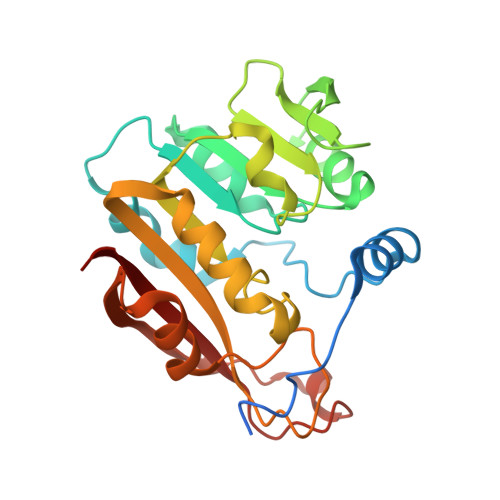 | |
UniProt | |||||
Find proteins for O55060 (Mus musculus) Explore O55060 Go to UniProtKB: O55060 | |||||
Entity Groups | |||||
| Sequence Clusters | 30% Identity50% Identity70% Identity90% Identity95% Identity100% Identity | ||||
| UniProt Group | O55060 | ||||
Sequence AnnotationsExpand | |||||
| |||||
| Ligands 1 Unique | |||||
|---|---|---|---|---|---|
| ID | Chains | Name / Formula / InChI Key | 2D Diagram | 3D Interactions | |
| SAH Query on SAH | C [auth A], D [auth B] | S-ADENOSYL-L-HOMOCYSTEINE C14 H20 N6 O5 S ZJUKTBDSGOFHSH-WFMPWKQPSA-N |  | ||
| Length ( Å ) | Angle ( ˚ ) |
|---|---|
| a = 62.988 | α = 90 |
| b = 70.554 | β = 115.7 |
| c = 72.593 | γ = 90 |
| Software Name | Purpose |
|---|---|
| SHELXL-97 | refinement |
| SCALEPACK | data scaling |
| PDB_EXTRACT | data extraction |
| DENZO | data reduction |
| MOLREP | phasing |
| REFMAC | refinement |
RCSB PDB (citation) is hosted by
RCSB PDB is a member of the

