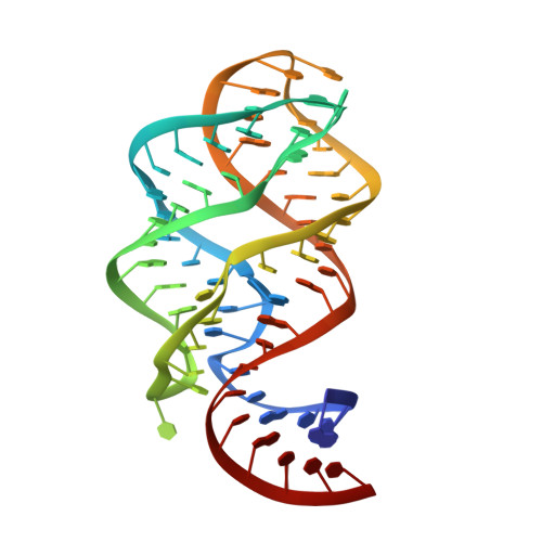
2G9C

Modified pyrimidines Specifically bind the purine riboswitch


Experimental Data Snapshot

  • Method: X-RAY DIFFRACTION
  • Resolution: 1.70 Å
  • R-Value Free: 0.242 
  • R-Value Work: 0.195 
  • R-Value Observed: 0.231 

wwPDB Validation   3D Report Full Report


This is version 1.4 of the entry. See complete history


Literature

Modified pyrimidines specifically bind the purine riboswitch.

Gilbert, S.D.Mediatore, S.J.Batey, R.T.

(2006) J Am Chem Soc 128: 14214-14215

  • DOI: https://doi.org/10.1021/ja063645t
  • Primary Citation of Related Structures:  
    2G9C

  • PubMed Abstract: 

    The purine riboswitch is a genetic regulatory element found in the 5'-untranslated regions of Gram-positive bacteria that regulates expression of the mRNA specifically in response to either guanine or adenine. We report that the adenine-responsive RNA element is also capable of specifically recognizing pyrimidine compounds bearing modifications at the 6- or 5,6-positions in a fashion similar to that of purine compounds. Using isothermal titration calorimetry and X-ray crystallography, the binding of these compounds is characterized.


  • Organizational Affiliation

    Department of Chemistry and Biochemistry, University of Colorado at Boulder, Campus Box 215, Boulder, Colorado 80309-0215, USA.


Macromolecules
Find similar nucleic acids by:  (by identity cutoff)  |  3D Structure
Entity ID: 1
MoleculeChains LengthOrganismImage
guanine riboswitch67N/A
Sequence Annotations
Expand
  • Reference Sequence
Small Molecules
Ligands 3 Unique
IDChains Name / Formula / InChI Key2D Diagram3D Interactions
NCO
Query on NCO

Download Ideal Coordinates CCD File 
C [auth A]
D [auth A]
E [auth A]
F [auth A]
G [auth A]
C [auth A],
D [auth A],
E [auth A],
F [auth A],
G [auth A],
H [auth A],
I [auth A],
J [auth A],
K [auth A],
L [auth A],
M [auth A],
N [auth A]
COBALT HEXAMMINE(III)
Co H18 N6
DYLMFCCYOUSRTK-UHFFFAOYSA-N
3AY
Query on 3AY

Download Ideal Coordinates CCD File 
O [auth A]PYRIMIDINE-2,4,6-TRIAMINE
C4 H7 N5
JTTIOYHBNXDJOD-UHFFFAOYSA-N
ACT
Query on ACT

Download Ideal Coordinates CCD File 
B [auth A]ACETATE ION
C2 H3 O2
QTBSBXVTEAMEQO-UHFFFAOYSA-M
Binding Affinity Annotations 
IDSourceBinding Affinity
3AY PDBBind:  2G9C Kd: 2000 (nM) from 1 assay(s)
Experimental Data & Validation

Experimental Data

  • Method: X-RAY DIFFRACTION
  • Resolution: 1.70 Å
  • R-Value Free: 0.242 
  • R-Value Work: 0.195 
  • R-Value Observed: 0.231 
  • Space Group: C 1 2 1
Unit Cell:
Length ( Å )Angle ( ˚ )
a = 131.99α = 90
b = 35.1β = 90.43
c = 41.8γ = 90
Software Package:
Software NamePurpose
CNSrefinement
BOSdata collection
d*TREKdata scaling
CNSphasing

Structure Validation

View Full Validation Report


Entry History 

Deposition Data

Revision History  (Full details and data files)

  • Version 1.0: 2006-11-21
    Type: Initial release
  • Version 1.1: 2008-05-01
    Changes: Version format compliance
  • Version 1.2: 2011-07-13
    Changes: Version format compliance
  • Version 1.3: 2017-10-18
    Changes: Refinement description
  • Version 1.4: 2024-02-14
    Changes: Data collection, Database references, Derived calculations