Light chain of botulinum neurotoxin serotype A: structural resolution of a catalytic intermediate.
Fu, Z., Chen, S., Baldwin, M.R., Boldt, G.E., Crawford, A., Janda, K.D., Barbieri, J.T., Kim, J.J.(2006) Biochemistry 45: 8903-8911
- PubMed: 16846233
- DOI: https://doi.org/10.1021/bi060786z
- Primary Citation of Related Structures:
2G7K, 2G7P, 2G7Q - PubMed Abstract:
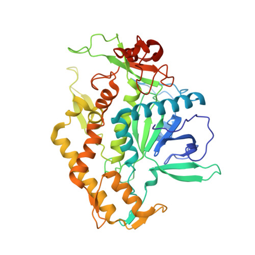
Botulinum neurotoxin serotype A (BoNT/A, 1296 residues) is a zinc metalloprotease that cleaves SNAP25 to inhibit the fusion of neurotransmitter-carrying vesicles to the plasma membrane of peripheral neurons. BoNT/A is a disulfide-linked di-chain protein composed of an N-terminal, thermolysin-like metalloprotease light chain domain (LC/A, 448 residues) and a C-terminal heavy chain domain (848 residues) that can be divided into two subdomains, a translocation subdomain and a receptor binding subdomain. LC/A cleaves SNAP25 between residues Gln197-Arg198 and, unlike thermolysin, recognizes an extended region of SNAP25 for cleavage. The structure of a recombinant LC/A (1-425) treated with EDTA (No-Zn LC/A) was determined. The overall structure of No-Zn LC/A is similar to that reported for the holotoxin, except that it lacks the Zn ion, indicating that the role of Zn is catalytic not structural. In addition, structures of a noncatalytic mutant LC/A (Arg362Ala/Tyr365Phe) complexed with and without an inhibitor, ArgHX, were determined. The overall structure and the active site conformation for the mutant are the same as wild type. When the inhibitor binds to the active site, the carbonyl and N-hydroxyl groups form a bidentate ligand to the Zn ion and the arginine moiety binds to Asp369, suggesting that the inhibitor-bound structure mimics a catalytic intermediate with the Arg moiety binding at the P1' site. Consistent with this model, mutation of Asp369 to Ala decreases the catalytic activity of LC/A by approximately 600-fold, and the residual activity is not inhibited by ArgHX. These results provide new information on the reaction mechanism and insight into the development of strategies for small molecule inhibitors of BoNTs.
Organizational Affiliation:
Department of Biochemistry, Medical College of Wisconsin, 8701 Watertown Plank Road, Milwaukee, Wisconsin 53226, USA.