2G6Z

Crystal structure of human DUSP5


Experimental Data Snapshot

  • Method: X-RAY DIFFRACTION
  • Resolution: 2.70 Å
  • R-Value Free: 0.291 
  • R-Value Work: 0.244 
  • R-Value Observed: 0.248 

wwPDB Validation   3D Report Full Report


This is version 1.2 of the entry. See complete history


Literature

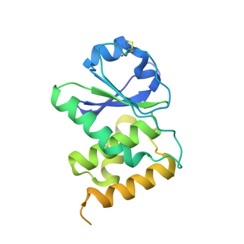
Crystal structure of the catalytic domain of human DUSP5, a dual specificity MAP kinase protein phosphatase

Jeong, D.G.Cho, Y.H.Yoon, T.S.Kim, J.H.Ryu, S.E.Kim, S.J.

(2007) Proteins 66: 253-258


Macromolecules
Find similar proteins by:  (by identity cutoff)  |  3D Structure
Entity ID: 1
MoleculeChains Sequence LengthOrganismDetailsImage
Dual specificity protein phosphatase 5
A, B, C
211Homo sapiensMutation(s): 0 
EC: 3.1.3.48 (PDB Primary Data), 3.1.3.16 (PDB Primary Data)
UniProt & NIH Common Fund Data Resources
Find proteins for Q16690 (Homo sapiens)
Explore Q16690 
Go to UniProtKB:  Q16690
PHAROS:  Q16690
GTEx:  ENSG00000138166 
Entity Groups  
Sequence Clusters30% Identity50% Identity70% Identity90% Identity95% Identity100% Identity
UniProt GroupQ16690
Sequence Annotations
Expand
  • Reference Sequence
Experimental Data & Validation

Experimental Data

  • Method: X-RAY DIFFRACTION
  • Resolution: 2.70 Å
  • R-Value Free: 0.291 
  • R-Value Work: 0.244 
  • R-Value Observed: 0.248 
  • Space Group: P 43 2 2
Unit Cell:
Length ( Å )Angle ( ˚ )
a = 92.712α = 90
b = 92.712β = 90
c = 165.213γ = 90
Software Package:
Software NamePurpose
MOSFLMdata reduction
SCALAdata scaling
EPMRphasing
CNSrefinement
CCP4data scaling

Structure Validation

View Full Validation Report


Entry History 

Deposition Data

Revision History  (Full details and data files)

  • Version 1.0: 2007-01-16
    Type: Initial release
  • Version 1.1: 2008-05-01
    Changes: Version format compliance
  • Version 1.2: 2011-07-13
    Changes: Version format compliance