2G50

The location of the allosteric amino acid binding site of muscle pyruvate kinase.


Experimental Data Snapshot

  • Method: X-RAY DIFFRACTION
  • Resolution: 1.65 Å
  • R-Value Free: 0.174 
  • R-Value Work: 0.148 
  • R-Value Observed: 0.149 

wwPDB Validation   3D Report Full Report


This is version 2.1 of the entry. See complete history


Literature

Differentiating a Ligand's Chemical Requirements for Allosteric Interactions from Those for Protein Binding. Phenylalanine Inhibition of Pyruvate Kinase.

Williams, R.Holyoak, T.McDonald, G.Gui, C.Fenton, A.W.

(2006) Biochemistry 45: 5421-5429

  • DOI: https://doi.org/10.1021/bi0524262
  • Primary Citation of Related Structures:  
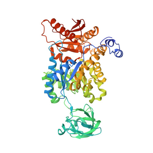
    2G50

  • PubMed Abstract: 

    The isoform of pyruvate kinase from brain and muscle of mammals (M(1)-PYK) is allosterically inhibited by phenylalanine. Initial observations in this model allosteric system indicate that Ala binds competitively with Phe, but elicits a minimal allosteric response. Thus, the allosteric ligand of this system must have requirements for eliciting an allosteric response in addition to the requirements for binding. Phe analogues have been used to dissect what chemical properties of Phe are responsible for eliciting the allosteric response. We first demonstrate that the l-2-aminopropanaldehyde substructure of the amino acid ligand is primarily responsible for binding to M(1)-PYK. Since the allosteric response to Ala is minimal and linear addition of methyl groups beyond the beta-carbon increase the magnitude of the allosteric response, we conclude that moieties beyond the beta-carbon are primarily responsible for allostery. Instead of an all-or-none mechanism of allostery, these findings support the idea that the bulk of the hydrophobic side chain, but not the aromatic nature, is the primary determinant of the magnitude of the observed allosteric inhibition. The use of these results to direct structural studies has resulted in a 1.65 A structure of M(1)-PYK with Ala bound. The coordination of Ala in the allosteric amino acid binding site confirms the binding role of the l-2-aminopropanaldehyde substructure of the ligand. Collectively, this study confirms that a ligand can have chemical regions specific for eliciting the allosteric signal in addition to the chemical regions necessary for binding.


  • Organizational Affiliation

    Department of Biochemistry and Molecular Biology, The University of Kansas Medical Center, Kansas City, Kansas 66160, USA.


Macromolecules
Find similar proteins by:  (by identity cutoff)  |  3D Structure
Entity ID: 1
MoleculeChains Sequence LengthOrganismDetailsImage
Pyruvate kinase isozymes M1/M2
A, B, C, D, E
A, B, C, D, E, F, G, H
530Oryctolagus cuniculusMutation(s): 0 
EC: 2.7.1.40
UniProt
Find proteins for P11974 (Oryctolagus cuniculus)
Explore P11974 
Go to UniProtKB:  P11974
Entity Groups  
Sequence Clusters30% Identity50% Identity70% Identity90% Identity95% Identity100% Identity
UniProt GroupP11974
Sequence Annotations
Expand
  • Reference Sequence
Small Molecules
Ligands 8 Unique
IDChains Name / Formula / InChI Key2D Diagram3D Interactions
ETE
Query on ETE

Download Ideal Coordinates CCD File 
DA [auth B]2-{2-[2-2-(METHOXY-ETHOXY)-ETHOXY]-ETHOXY}-ETHANOL
C9 H20 O5
ZNYRFEPBTVGZDN-UHFFFAOYSA-N
GOL
Query on GOL

Download Ideal Coordinates CCD File 
AC [auth F]
BC [auth F]
CB [auth D]
CC [auth F]
CD [auth H]
AC [auth F],
BC [auth F],
CB [auth D],
CC [auth F],
CD [auth H],
DB [auth D],
DC [auth F],
DD [auth H],
EA [auth B],
EC [auth F],
ED [auth H],
FA [auth B],
FC [auth F],
FD [auth H],
GA [auth B],
NC [auth G],
OA [auth C],
OC [auth G],
PA [auth C],
PC [auth G],
QA [auth C],
QB [auth E],
R [auth A],
RA [auth C],
RB [auth E],
S [auth A],
SB [auth E],
T [auth A]
GLYCEROL
C3 H8 O3
PEDCQBHIVMGVHV-UHFFFAOYSA-N
ALA
Query on ALA

Download Ideal Coordinates CCD File 
IB [auth E]
JC [auth G]
KA [auth C]
M [auth A]
UC [auth H]
IB [auth E],
JC [auth G],
KA [auth C],
M [auth A],
UC [auth H],
WA [auth D],
WB [auth F],
X [auth B]
ALANINE
C3 H7 N O2
QNAYBMKLOCPYGJ-REOHCLBHSA-N
PYR
Query on PYR

Download Ideal Coordinates CCD File 
JB [auth E]
KC [auth G]
LA [auth C]
N [auth A]
VC [auth H]
JB [auth E],
KC [auth G],
LA [auth C],
N [auth A],
VC [auth H],
XA [auth D],
XB [auth F],
Y [auth B]
PYRUVIC ACID
C3 H4 O3
LCTONWCANYUPML-UHFFFAOYSA-N
EDO
Query on EDO

Download Ideal Coordinates CCD File 
AA [auth B]
AB [auth D]
AD [auth H]
BA [auth B]
BB [auth D]
AA [auth B],
AB [auth D],
AD [auth H],
BA [auth B],
BB [auth D],
BD [auth H],
CA [auth B],
KB [auth E],
LB [auth E],
LC [auth G],
MA [auth C],
MB [auth E],
MC [auth G],
NA [auth C],
NB [auth E],
O [auth A],
OB [auth E],
P [auth A],
PB [auth E],
Q [auth A],
WC [auth H],
XC [auth H],
YA [auth D],
YB [auth F],
YC [auth H],
Z [auth B],
ZA [auth D],
ZB [auth F],
ZC [auth H]
1,2-ETHANEDIOL
C2 H6 O2
LYCAIKOWRPUZTN-UHFFFAOYSA-N
MN
Query on MN

Download Ideal Coordinates CCD File 
EB [auth E]
GC [auth G]
HA [auth C]
I [auth A]
QC [auth H]
EB [auth E],
GC [auth G],
HA [auth C],
I [auth A],
QC [auth H],
SA [auth D],
TB [auth F],
U [auth B]
MANGANESE (II) ION
Mn
WAEMQWOKJMHJLA-UHFFFAOYSA-N
K
Query on K

Download Ideal Coordinates CCD File 
FB [auth E]
HC [auth G]
IA [auth C]
J [auth A]
RC [auth H]
FB [auth E],
HC [auth G],
IA [auth C],
J [auth A],
RC [auth H],
TA [auth D],
UB [auth F],
V [auth B]
POTASSIUM ION
K
NPYPAHLBTDXSSS-UHFFFAOYSA-N
NA
Query on NA

Download Ideal Coordinates CCD File 
GB [auth E]
HB [auth E]
IC [auth G]
JA [auth C]
K [auth A]
GB [auth E],
HB [auth E],
IC [auth G],
JA [auth C],
K [auth A],
L [auth A],
SC [auth H],
TC [auth H],
UA [auth D],
VA [auth D],
VB [auth F],
W [auth B]
SODIUM ION
Na
FKNQFGJONOIPTF-UHFFFAOYSA-N
Experimental Data & Validation

Experimental Data

  • Method: X-RAY DIFFRACTION
  • Resolution: 1.65 Å
  • R-Value Free: 0.174 
  • R-Value Work: 0.148 
  • R-Value Observed: 0.149 
  • Space Group: P 1
Unit Cell:
Length ( Å )Angle ( ˚ )
a = 82.626α = 95.15
b = 109.014β = 93.45
c = 144.463γ = 112.26
Software Package:
Software NamePurpose
DENZOdata reduction
SCALEPACKdata scaling
REFMACrefinement
PDB_EXTRACTdata extraction
MOLREPphasing

Structure Validation

View Full Validation Report


Entry History 

Deposition Data

Revision History  (Full details and data files)

  • Version 1.0: 2006-05-09
    Type: Initial release
  • Version 1.1: 2008-05-01
    Changes: Version format compliance
  • Version 1.2: 2011-07-13
    Changes: Advisory, Refinement description, Version format compliance
  • Version 1.3: 2017-10-18
    Changes: Refinement description
  • Version 2.0: 2023-11-15
    Changes: Atomic model, Data collection, Database references, Derived calculations
  • Version 2.1: 2024-04-03
    Changes: Refinement description