2FYF

Structure of a putative phosphoserine aminotransferase from Mycobacterium Tuberculosis


Experimental Data Snapshot

  • Method: X-RAY DIFFRACTION
  • Resolution: 1.50 Å
  • R-Value Free: 0.166 
  • R-Value Work: 0.144 
  • R-Value Observed: 0.145 

wwPDB Validation   3D Report Full Report


Ligand Structure Quality Assessment 


This is version 1.5 of the entry. See complete history


Literature

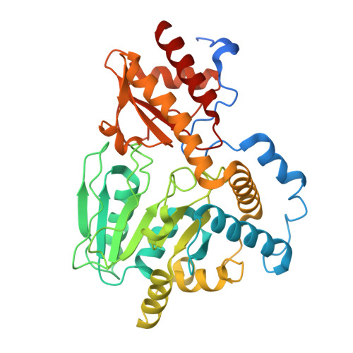
Structure of phosphoserine aminotransferase from Mycobacterium tuberculosis.

Coulibaly, F.Lassalle, E.Baker, H.M.Baker, E.N.

(2012) Acta Crystallogr D Biol Crystallogr 68: 553-563

  • DOI: https://doi.org/10.1107/S0907444912004829
  • Primary Citation of Related Structures:  
    2FYF, 3VOM

  • PubMed Abstract: 

    Mycobacterium tuberculosis (Mtb), the causative agent of TB, remains a serious world health problem owing to limitations of the available drugs and the emergence of resistant strains. In this context, key biosynthetic enzymes from Mtb are attractive targets for the development of new therapeutic drugs. Here, the 1.5 Å resolution crystal structure of Mtb phosphoserine aminotransferase (MtbPSAT) in complex with its cofactor, pyridoxal 5'-phosphate (PLP), is reported. MtbPSAT is an essential enzyme in the biosynthesis of serine and in pathways of one-carbon metabolism. The structure shows that although the Mtb enzyme differs substantially in sequence from other PSAT enzymes, its fold is conserved and its PLP-binding site is virtually identical. Structural comparisons suggest that this site remains unchanged throughout the catalytic cycle. On the other hand, PSAT enzymes are obligate dimers in which the two active sites are located in the dimer interface and distinct differences in the MtbPSAT dimer are noted. These impact on the substrate-binding region and access channel and suggest options for the development of selective inhibitors.


  • Organizational Affiliation

    Maurice Wilkins Centre for Molecular Biodiscovery and School of Biological Sciences, University of Auckland, Private Bag 92019, Auckland, New Zealand.


Macromolecules
Find similar proteins by:  (by identity cutoff)  |  3D Structure
Entity ID: 1
MoleculeChains Sequence LengthOrganismDetailsImage
phosphoserine aminotransferase
A, B
398Mycobacterium tuberculosis H37RvMutation(s): 0 
Gene Names: serC
EC: 2.6.1.52
UniProt
Find proteins for P9WQ73 (Mycobacterium tuberculosis (strain ATCC 25618 / H37Rv))
Explore P9WQ73 
Go to UniProtKB:  P9WQ73
Entity Groups  
Sequence Clusters30% Identity50% Identity70% Identity90% Identity95% Identity100% Identity
UniProt GroupP9WQ73
Sequence Annotations
Expand
  • Reference Sequence
Small Molecules
Ligands 4 Unique
IDChains Name / Formula / InChI Key2D Diagram3D Interactions
PC4
Query on PC4

Download Ideal Coordinates CCD File 
D [auth A]
F [auth A]
K [auth B]
L [auth B]
N [auth B]
D [auth A],
F [auth A],
K [auth B],
L [auth B],
N [auth B],
O [auth B]
TETRACHLOROPLATINATE(II)
Cl4 Pt
SVZRVTAEYVVVPM-UHFFFAOYSA-J
PLP
Query on PLP

Download Ideal Coordinates CCD File 
E [auth A],
M [auth B]
PYRIDOXAL-5'-PHOSPHATE
C8 H10 N O6 P
NGVDGCNFYWLIFO-UHFFFAOYSA-N
SO4
Query on SO4

Download Ideal Coordinates CCD File 
C [auth A],
J [auth B]
SULFATE ION
O4 S
QAOWNCQODCNURD-UHFFFAOYSA-L
GOL
Query on GOL

Download Ideal Coordinates CCD File 
G [auth A]
H [auth A]
I [auth A]
P [auth B]
Q [auth B]
G [auth A],
H [auth A],
I [auth A],
P [auth B],
Q [auth B],
R [auth B],
S [auth B]
GLYCEROL
C3 H8 O3
PEDCQBHIVMGVHV-UHFFFAOYSA-N
Experimental Data & Validation

Experimental Data

  • Method: X-RAY DIFFRACTION
  • Resolution: 1.50 Å
  • R-Value Free: 0.166 
  • R-Value Work: 0.144 
  • R-Value Observed: 0.145 
  • Space Group: P 21 21 21
Unit Cell:
Length ( Å )Angle ( ˚ )
a = 77.478α = 90
b = 94.056β = 90
c = 101.063γ = 90
Software Package:
Software NamePurpose
DENZOdata reduction
SCALEPACKdata scaling
SHARPphasing
SOLOMONphasing
REFMACrefinement
PDB_EXTRACTdata extraction

Structure Validation

View Full Validation Report


Ligand Structure Quality Assessment 


Entry History 

Deposition Data

Revision History  (Full details and data files)

  • Version 1.0: 2007-01-16
    Type: Initial release
  • Version 1.1: 2008-05-01
    Changes: Version format compliance
  • Version 1.2: 2011-07-13
    Changes: Advisory, Source and taxonomy, Version format compliance
  • Version 1.3: 2012-06-13
    Changes: Database references
  • Version 1.4: 2017-10-18
    Changes: Advisory, Refinement description
  • Version 1.5: 2024-02-14
    Changes: Advisory, Data collection, Database references, Derived calculations