2FX3

Crystal Structure Determination of E. coli Elongation Factor, Tu using a Twinned Data Set


Experimental Data Snapshot

  • Method: X-RAY DIFFRACTION
  • Resolution: 3.40 Å
  • R-Value Free: 0.262 
  • R-Value Work: 0.197 

wwPDB Validation   3D Report Full Report


Ligand Structure Quality Assessment 


This is version 1.3 of the entry. See complete history


Literature

Solving the structure of Escherichia coli elongation factor Tu using a twinned data set.

Heffron, S.E.Moeller, R.Jurnak, F.

(2006) Acta Crystallogr D Biol Crystallogr 62: 433-438

  • DOI: https://doi.org/10.1107/S0907444906004021
  • Primary Citation of Related Structures:  
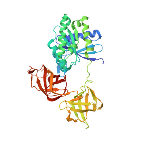
    2FX3

  • PubMed Abstract: 

    Escherichia coli elongation factor Tu-GDP (EF-Tu-GDP) was crystallized in the presence of novel inhibitors. The only crystals which could be grown were epitaxially as well as merohedrally twinned, highly mosaic and diffracted to a resolution of 3.4 A in space group P3(1)21, with unit-cell parameters a = b = 69.55, c = 169.44 A, alpha = beta = 90, gamma = 120 degrees . To determine whether an inhibitor was present in the crystal, a poor-quality X-ray diffraction data set had to be processed. The three-dimensional structure was ultimately solved and the original question answered. The results also reveal a new type of dimer packing for EF-Tu-GDP.


  • Organizational Affiliation

    University of California, Irvine, USA.


Macromolecules
Find similar proteins by:  (by identity cutoff)  |  3D Structure
Entity ID: 1
MoleculeChains Sequence LengthOrganismDetailsImage
Elongation factor Tu393Escherichia coliMutation(s): 0 
UniProt
Find proteins for P0CE47 (Escherichia coli (strain K12))
Explore P0CE47 
Go to UniProtKB:  P0CE47
Entity Groups  
Sequence Clusters30% Identity50% Identity70% Identity90% Identity95% Identity100% Identity
UniProt GroupP0CE47
Sequence Annotations
Expand
  • Reference Sequence
Small Molecules
Ligands 2 Unique
IDChains Name / Formula / InChI Key2D Diagram3D Interactions
GDP
Query on GDP

Download Ideal Coordinates CCD File 
C [auth A]GUANOSINE-5'-DIPHOSPHATE
C10 H15 N5 O11 P2
QGWNDRXFNXRZMB-UUOKFMHZSA-N
MG
Query on MG

Download Ideal Coordinates CCD File 
B [auth A]MAGNESIUM ION
Mg
JLVVSXFLKOJNIY-UHFFFAOYSA-N
Experimental Data & Validation

Experimental Data

  • Method: X-RAY DIFFRACTION
  • Resolution: 3.40 Å
  • R-Value Free: 0.262 
  • R-Value Work: 0.197 
  • Space Group: P 31 2 1
Unit Cell:
Length ( Å )Angle ( ˚ )
a = 69.55α = 90
b = 69.55β = 90
c = 169.44γ = 120
Software Package:
Software NamePurpose
SCALAdata scaling
PHASERphasing
CNSrefinement
PDB_EXTRACTdata extraction
MOSFLMdata reduction
CCP4data scaling

Structure Validation

View Full Validation Report


Ligand Structure Quality Assessment 


Entry History 

Deposition Data

Revision History  (Full details and data files)

  • Version 1.0: 2006-03-28
    Type: Initial release
  • Version 1.1: 2008-05-01
    Changes: Version format compliance
  • Version 1.2: 2011-07-13
    Changes: Version format compliance
  • Version 1.3: 2023-08-30
    Changes: Data collection, Database references, Derived calculations, Refinement description