2FTL

Crystal structure of trypsin complexed with BPTI at 100K


Experimental Data Snapshot

  • Method: X-RAY DIFFRACTION
  • Resolution: 1.62 Å
  • R-Value Free: 0.227 
  • R-Value Work: 0.214 
  • R-Value Observed: 0.215 

wwPDB Validation   3D Report Full Report


This is version 1.4 of the entry. See complete history


Literature

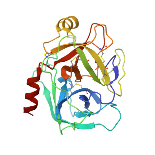
Rigidification of a Flexible Protease Inhibitor Variant upon Binding to Trypsin.

Hanson, W.M.Domek, G.J.Horvath, M.P.Goldenberg, D.P.

(2007) J Mol Biol 366: 230-243

  • DOI: https://doi.org/10.1016/j.jmb.2006.11.003
  • Primary Citation of Related Structures:  
    2FTL, 2FTM

  • PubMed Abstract: 

    The Tyr35-->Gly replacement in bovine pancreatic trypsin inhibitor (BPTI) has previously been shown to dramatically enhance the flexibility of the trypsin-binding region of the free inhibitor and to destabilize the interaction with the protease by about 3 kcal/mol. The effects of this replacement on the enzyme-inhibitor interaction were further studied here by X-ray crystallography and isothermal titration calorimetry (ITC). The co-crystal structure of Y35G BPTI bound to trypsin was determined using 1.65 A resolution X-ray diffraction data collected from cryopreserved crystals, and a new structure of the complex with wild-type BPTI under the same conditions was determined using 1.62 A data. These structures reveal that, in contrast to the free protein, Y35G BPTI adopts a conformation nearly identical with that of the wild-type protein, with a water-filled cavity in place of the missing Tyr side-chain. The crystallographic temperature factors for the two complexes indicate that the mutant inhibitor is nearly as rigid as the wild-type protein when bound to trypsin. Calorimetric measurements show that the change in enthalpy upon dissociation of the complex is 2.5 kcal/mol less favorable for the complex containing Y35G BPTI than for the complex with the wild-type inhibitor. Thus, the destabilization of the complex resulting from the Y35G replacement is due to a more favorable change in entropy upon dissociation. The heat capacity changes for dissociation of the mutant and wild-type complexes were very similar, suggesting that the entropic effects probably do not arise from solvation effects, but are more likely due to an increase in protein conformational entropy upon dissociation of the mutant inhibitor. These results define the biophysical role of a highly conserved core residue located outside of a protein-binding interface, demonstrating that Tyr35 has little impact on the trypsin-bound BPTI structure and acts primarily to define the structure of the free protein so as to maximize binding affinity.


  • Organizational Affiliation

    Department of Biology, University of Utah, 257 South 1400 East, Salt Lake City, UT 84112-0840, USA.


Macromolecules
Find similar proteins by:  (by identity cutoff)  |  3D Structure
Entity ID: 1
MoleculeChains Sequence LengthOrganismDetailsImage
Cationic trypsinA [auth E]223Bos taurusMutation(s): 0 
EC: 3.4.21.4
UniProt
Find proteins for P00760 (Bos taurus)
Explore P00760 
Go to UniProtKB:  P00760
Entity Groups  
Sequence Clusters30% Identity50% Identity70% Identity90% Identity95% Identity100% Identity
UniProt GroupP00760
Sequence Annotations
Expand
  • Reference Sequence
Find similar proteins by:  (by identity cutoff)  |  3D Structure
Entity ID: 2
MoleculeChains Sequence LengthOrganismDetailsImage
Pancreatic trypsin inhibitorB [auth I]58Bos taurusMutation(s): 0 
UniProt
Find proteins for P00974 (Bos taurus)
Explore P00974 
Go to UniProtKB:  P00974
Entity Groups  
Sequence Clusters30% Identity50% Identity70% Identity90% Identity95% Identity100% Identity
UniProt GroupP00974
Sequence Annotations
Expand
  • Reference Sequence
Small Molecules
Ligands 4 Unique
IDChains Name / Formula / InChI Key2D Diagram3D Interactions
SO4
Query on SO4

Download Ideal Coordinates CCD File 
E
F [auth E]
G [auth E]
H [auth E]
P [auth I]
E,
F [auth E],
G [auth E],
H [auth E],
P [auth I],
Q [auth I],
R [auth I],
S [auth I]
SULFATE ION
O4 S
QAOWNCQODCNURD-UHFFFAOYSA-L
EDO
Query on EDO

Download Ideal Coordinates CCD File 
I [auth E]
J [auth E]
K [auth E]
L [auth E]
M [auth E]
I [auth E],
J [auth E],
K [auth E],
L [auth E],
M [auth E],
N [auth E]
1,2-ETHANEDIOL
C2 H6 O2
LYCAIKOWRPUZTN-UHFFFAOYSA-N
CA
Query on CA

Download Ideal Coordinates CCD File 
D [auth E],
O [auth I]
CALCIUM ION
Ca
BHPQYMZQTOCNFJ-UHFFFAOYSA-N
NA
Query on NA

Download Ideal Coordinates CCD File 
C [auth E]SODIUM ION
Na
FKNQFGJONOIPTF-UHFFFAOYSA-N
Experimental Data & Validation

Experimental Data

  • Method: X-RAY DIFFRACTION
  • Resolution: 1.62 Å
  • R-Value Free: 0.227 
  • R-Value Work: 0.214 
  • R-Value Observed: 0.215 
  • Space Group: I 2 2 2
Unit Cell:
Length ( Å )Angle ( ˚ )
a = 74.726α = 90
b = 81.906β = 90
c = 124.261γ = 90
Software Package:
Software NamePurpose
HKL-2000data collection
SCALEPACKdata scaling
CNSrefinement
HKL-2000data reduction
CNSphasing

Structure Validation

View Full Validation Report


Entry History 

Deposition Data

Revision History  (Full details and data files)

  • Version 1.0: 2006-02-14
    Type: Initial release
  • Version 1.1: 2008-05-01
    Changes: Version format compliance
  • Version 1.2: 2011-07-13
    Changes: Version format compliance
  • Version 1.3: 2011-07-27
    Changes: Database references, Derived calculations, Non-polymer description
  • Version 1.4: 2023-08-30
    Changes: Advisory, Data collection, Database references, Derived calculations, Refinement description