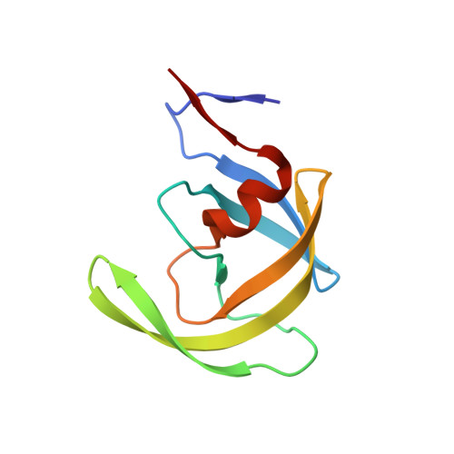
2FLE

Structural analysis of asymmetric inhibitor bound to the HIV-1 Protease V82A mutant


Experimental Data Snapshot

  • Method: X-RAY DIFFRACTION
  • Resolution: 1.90 Å
  • R-Value Free: 0.251 
  • R-Value Work: 0.199 
  • R-Value Observed: 0.207 

wwPDB Validation   3D Report Full Report


Ligand Structure Quality Assessment 


This is version 1.5 of the entry. See complete history


Literature

Design, synthesis, evaluation, and crystallographic-based structural studies of HIV-1 protease inhibitors with reduced response to the V82A mutation.

Clemente, J.C.Robbins, A.Grana, P.Paleo, M.R.Correa, J.F.Villaverde, M.C.Sardina, F.J.Govindasamy, L.Agbandje-McKenna, M.McKenna, R.Dunn, B.M.Sussman, F.

(2008) J Med Chem 51: 852-860

  • DOI: https://doi.org/10.1021/jm701170f
  • Primary Citation of Related Structures:  
    2FLE

  • PubMed Abstract: 

    In our quest for HIV-1 protease inhibitors that are not affected by the V82A resistance mutation, we have synthesized and tested a second generation set of C2-symmetric HIV-1 protease inhibitors that contain a cyclohexane group at P1 and/or P1'. The binding affinity results indicate that these compounds have an improved response to the appearance of the V82A mutation than the parent compound. The X-ray structure of one of these compounds with the V82A HIV-1 PR variant provides the structural rationale for the better resistance profile of these compounds. Moreover, scrutiny of the X-ray structure suggests that the ring of the Cha side chain might be in a boat rather than in the chair conformation, a result supported by molecular dynamics simulations.


  • Organizational Affiliation

    Department of Biochemistry and Molecular Biology, University of Florida College of Medicine, Gainesville, Florida 32610, USA.


Macromolecules
Find similar proteins by:  (by identity cutoff)  |  3D Structure
Entity ID: 1
MoleculeChains Sequence LengthOrganismDetailsImage
pol protein
A, B
99Human immunodeficiency virus 1Mutation(s): 1 
UniProt
Find proteins for Q9J2R0 (Human immunodeficiency virus 1)
Explore Q9J2R0 
Go to UniProtKB:  Q9J2R0
Entity Groups  
Sequence Clusters30% Identity50% Identity70% Identity90% Identity95% Identity100% Identity
UniProt GroupQ9J2R0
Sequence Annotations
Expand
  • Reference Sequence
Small Molecules
Ligands 2 Unique
IDChains Name / Formula / InChI Key2D Diagram3D Interactions
AI
Query on AI

Download Ideal Coordinates CCD File 
D [auth B](2S,2'S)-N,N'-[(2S,3S,4S,5S)-1-CYCLOHEXYL-3,4-DIHYDROXY-6-PHENYLHEXANE-2,5-DIYL]BIS[3-METHYL-2-({[METHYL(PYRIDIN-2-YLMETHYL)AMINO]CARBONYL}AMINO)BUTANAMIDE]
C44 H64 N8 O6
JQIFSYRTTKZQMY-UNHORJANSA-N
GOL
Query on GOL

Download Ideal Coordinates CCD File 
C [auth A]GLYCEROL
C3 H8 O3
PEDCQBHIVMGVHV-UHFFFAOYSA-N
Binding Affinity Annotations 
IDSourceBinding Affinity
AI BindingDB:  2FLE Ki: min: 3.2, max: 14 (nM) from 2 assay(s)
PDBBind:  2FLE Ki: 14 (nM) from 1 assay(s)
Binding MOAD:  2FLE Ki: 14 (nM) from 1 assay(s)
Experimental Data & Validation

Experimental Data

  • Method: X-RAY DIFFRACTION
  • Resolution: 1.90 Å
  • R-Value Free: 0.251 
  • R-Value Work: 0.199 
  • R-Value Observed: 0.207 
  • Space Group: P 61
Unit Cell:
Length ( Å )Angle ( ˚ )
a = 62.195α = 90
b = 62.195β = 90
c = 82.752γ = 120
Software Package:
Software NamePurpose
DENZOdata reduction
SCALEPACKdata scaling
CNSrefinement
PDB_EXTRACTdata extraction
HKL-2000data reduction
CNSphasing

Structure Validation

View Full Validation Report


Ligand Structure Quality Assessment 


Entry History 

Deposition Data

Revision History  (Full details and data files)

  • Version 1.0: 2007-01-16
    Type: Initial release
  • Version 1.1: 2008-05-01
    Changes: Version format compliance
  • Version 1.2: 2011-07-13
    Changes: Non-polymer description, Version format compliance
  • Version 1.3: 2017-10-18
    Changes: Refinement description
  • Version 1.4: 2021-10-20
    Changes: Database references, Derived calculations
  • Version 1.5: 2023-08-30
    Changes: Data collection, Refinement description