2FID

Crystal Structure of a Bovine Rabex-5 fragment complexed with ubiquitin


Experimental Data Snapshot

  • Method: X-RAY DIFFRACTION
  • Resolution: 2.80 Å
  • R-Value Free: 0.268 
  • R-Value Work: 0.211 
  • R-Value Observed: 0.217 

wwPDB Validation   3D Report Full Report


This is version 1.3 of the entry. See complete history


Literature

Structural basis for ubiquitin recognition and autoubiquitination by Rabex-5

Lee, S.Tsai, Y.C.Mattera, R.Smith, W.J.Kostelansky, M.S.Weissman, A.M.Bonifacino, J.S.Hurley, J.H.

(2006) Nat Struct Mol Biol 13: 264-271

  • DOI: https://doi.org/10.1038/nsmb1064
  • Primary Citation of Related Structures:  
    2FID, 2FIF

  • PubMed Abstract: 

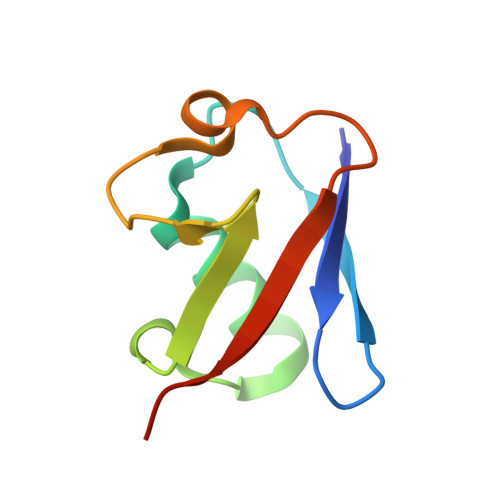
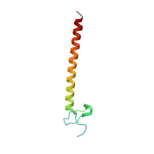
    Rabex-5 is an exchange factor for Rab5, a master regulator of endosomal trafficking. Rabex-5 binds monoubiquitin, undergoes covalent ubiquitination and contains an intrinsic ubiquitin ligase activity, all of which require an N-terminal A20 zinc finger followed immediately by a helix. The structure of the N-terminal portion of Rabex-5 bound to ubiquitin at 2.5-A resolution shows that Rabex-5-ubiquitin interactions occur at two sites. The first site is a new type of ubiquitin-binding domain, an inverted ubiquitin-interacting motif, which binds with approximately 29-microM affinity to the canonical Ile44 hydrophobic patch on ubiquitin. The second is a diaromatic patch on the A20 zinc finger, which binds with approximately 22-microM affinity to a polar region centered on Asp58 of ubiquitin. The A20 zinc-finger diaromatic patch mediates ubiquitin-ligase activity by directly recruiting a ubiquitin-loaded ubiquitin-conjugating enzyme.


  • Organizational Affiliation

    Laboratory of Molecular Biology, National Institute of Diabetes and Digestive and Kidney Diseases, US National Institutes of Health (NIH), Bethesda, Maryland 20892, USA.


Macromolecules
Find similar proteins by:  (by identity cutoff)  |  3D Structure
Entity ID: 1
MoleculeChains Sequence LengthOrganismDetailsImage
Ubiquitin76Bos taurusMutation(s): 0 
UniProt
Find proteins for P0CH28 (Bos taurus)
Explore P0CH28 
Go to UniProtKB:  P0CH28
Entity Groups  
Sequence Clusters30% Identity50% Identity70% Identity90% Identity95% Identity100% Identity
UniProt GroupP0CH28
Sequence Annotations
Expand
  • Reference Sequence
Find similar proteins by:  (by identity cutoff)  |  3D Structure
Entity ID: 2
MoleculeChains Sequence LengthOrganismDetailsImage
Rab5 GDP/GTP exchange factor70Bos taurusMutation(s): 0 
Gene Names: RABGEF1RABEX5
UniProt
Find proteins for O18973 (Bos taurus)
Explore O18973 
Go to UniProtKB:  O18973
Entity Groups  
Sequence Clusters30% Identity50% Identity70% Identity90% Identity95% Identity100% Identity
UniProt GroupO18973
Sequence Annotations
Expand
  • Reference Sequence
Small Molecules
Ligands 1 Unique
IDChains Name / Formula / InChI Key2D Diagram3D Interactions
ZN
Query on ZN

Download Ideal Coordinates CCD File 
C [auth B]ZINC ION
Zn
PTFCDOFLOPIGGS-UHFFFAOYSA-N
Experimental Data & Validation

Experimental Data

  • Method: X-RAY DIFFRACTION
  • Resolution: 2.80 Å
  • R-Value Free: 0.268 
  • R-Value Work: 0.211 
  • R-Value Observed: 0.217 
  • Space Group: P 61
Unit Cell:
Length ( Å )Angle ( ˚ )
a = 81.967α = 90
b = 81.967β = 90
c = 54.443γ = 120
Software Package:
Software NamePurpose
REFMACrefinement
HKL-2000data reduction
SCALEPACKdata scaling
SOLVEphasing

Structure Validation

View Full Validation Report


Entry History 

Deposition Data

Revision History  (Full details and data files)

  • Version 1.0: 2006-02-07
    Type: Initial release
  • Version 1.1: 2008-05-01
    Changes: Version format compliance
  • Version 1.2: 2011-07-13
    Changes: Advisory, Version format compliance
  • Version 1.3: 2024-02-14
    Changes: Data collection, Database references, Derived calculations